
Recent Studies
Manipulation of Light with Minerals (together with Lia Addadi and Dan Oron)
Ever since the study of the lens arrays in the brittlestar that started in our group (Aizenberg et al., 2001), we have been fascinated by the possibility that light manipulation by biogenic minerals is a widespread phenomenon, not only to produce structural colors, but also possibly on a huge scale in the plant kingdom that after all, “makes its living” by manipulating light. We have elucidated the structural basis by which fish and spiders produce metallic lusters based on ordered stacks of extremely thin arrays of guanine crystals (Levy-Lior et al., 2010). In plants we have shown that certain mineral bodies in leaves referred to as cystoliths (and composed of various forms of ACC stabilized by silicates) scatter light deep into the leaf and in this way make many more photons available for photosynthesis (Gal et al., 2012).
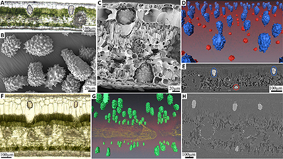 |
The anatomy of F. microcarpa cystoliths (A-E) and C. illinoinensis calcium oxalate druses (F-J). A+F) Light microscopy images of fresh, 50m thick leaf slices. The green mesophyll is perforated by the transparent biominerals; B+G) Scanning electron microscope images of extracted cystoliths (B) and druses (G); C+H) Scanning electron microscope images of cross-sections of critical-point-dried leaves showing cystoliths (C) and druses (H) in their anatomical location; D+I) Perspective views of 3D surface rendered CT tomograms. The minerals are artificially colored. In (D): blue – upper epidermis cystoliths, red - lower epidermis cystolith. In (I): magenta - calcium oxalate druses (see also movies SM1+2). E+J) Slices of reconstructed CT tomograms. Minerals, as well as other anatomical structures, are visible. Color coded ellipses, as in (D)+(I), indicate biominerals. Figure is from (Gal et al., 2012). |