Research

Cholinergic Mechanisms - Structure/Function

For more than thirty years, I have had the rare opportunity to be able to work closely with Prof. Israel Silman, of the Neurobiology Dept, on studying proteins involed in neural transmission via cholinergic mechanisms, Our work has been focused on studies of the synaptic enzyme, acetylcholinesterase (AChE), whose principal role is termination of synaptic transmission at cholinergic synapses by rapid hydrolysis of the neurotransmitter, acetylcholine. For over 50 years, AChE has been the subject of intense interest for enzymologists, pharmacologists and toxicologists, due to its extremely high catalytic activity, a requirement for its biological activity, and since it is the target of the first generation of anti-Alzheimer (AD) drugs and of potent nerve agents and insecticides.

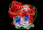
From the onset, our joint objective was determination of the 3D structure of AChE so as to understand its mechanism of action and permit structure-based drug design. Although attempts started in the ’60s, no crystals suitable for 3D structure determination were obtained prior to our pioneering work. We developed a novel, mild approach to solubilization of membrane-bound AChE from Torpedo californica electric organ (TcAChE), followed by its crystallization, and then by solution of its 3D structure. Our publication of the structure, as a full paper in Science, has been cited over 2,450 times, and many ensuing papers hundreds of times. We subsequently solved and published two additional important AChE structures, those of human and Drosophila AChE. Concomitantly, we solved the structures of more than 40 complexes and conjugates of AChE with a broad repertoire of drugs, toxins and other ligands. These included essentially all the 1st generation anti-AD drugs, several of the major nerve agents, insecticides, transition-state analogs and a snake venom toxin. These structural data provide an essential reservoir of information for generation of novel lead drugs. We have published over 170 joint papers.

Proteins from extreme environments

Most proteins function within a narrow range of salt concentration. However, in a project carried out in close collaboration with Prof. Ada Zamir, Dept of Biomolecular Sciences, studies of halotolerant Dunaliella salina revealed a new class of proteins - halotolerant proteins - that can function in the entire range of salinity (0 to 4 M NaCl). In order to get an insight into molecular details of how do these proteins take up the challenge to function in the varying salt concentrations, we have determined the first crystal structure of a halotolerant protein - a 30kDa Carbonic anhydrase.

The crystal structure coupled with the series of site-directed mutants will aid in for the better understanding of: what is the unique nature of protein - solvent and weak interactions for these class of proteins? What is the structural principle that confers the additional stability of halotolerance. How do halotolerant enzyme protect themselves from anionic/ cationic inhibition? Could this understanding have biotechnological applications such as engineering the enzymes to function in special solvents for industrial or environmental use?

AChE - Review

This review was write by Prof. Kurt Giles, while he was a post-doc in the Silman/Sussman lab in 1996-99.

Dale's momentous 1914 paper, in which he differentiated between the muscarine- and nicotine- like actions of choline esters on different tissues, proposed: "it seems not improbable that an esterase contributes to the removal of [acetylcholine] from the circulation". This hypothesis was based on observations of the inactivation of acetylcholine (ACh) injected into cats. However, it wasn't until 1926 that Loewi and Navratil, working on isolated frog's hearts, experimentally demonstrated its existence by inhibition with physostigmine (eserine), thus prolonging the effect of administered ACh. In 1932 Stedman et al. prepared a crude extract of an ACh-splitting enzyme from horse serum, which they called "cholinesterase."

Proteopedia - Life in 3D

Proteopedia is a collaborative, 3D web-encyclopedia of protein, nucleic acid and otherbiomolecule structures. Created as a means for communicating biomolecule structures to a diverse scientific audience, Proteopedia  presents structural annotation in an intuitive, interactive format and allows members of the scientific community to easily contribute their own annotations. There are  ~450,000 pages in Proteopedia contributed by more than 4,800 users from over 60 countries worldwide, with many pages translated into over a dozen different languages.

Acetylcholinesterase-Interacting Anti-Alzheimer's Drugs

While advances in medicine have increased the life expectancy of the overall population, the number of people affected by age related pathologies has also increased. The challenge facing physicians and scientists, then, is not just to help people live longer, but to be healthy and functional during their extended lifetimes. One of the most common afflictions of old age is senility, or dementia, which is the inexorable decline of cognitive ability, beginning with memory loss, and ending with complete helplessness. The most common cause of dementia is Alzheimer's disease (AD), affecting about 10% of the population over 65, 25% of the people over 75, and up to 45% of the people over 80. It is estimated that there are currently 4.5 million people suffering from Alzheimer's disease in the US alone.

Proteomic signatures: AA & peptide compositions differentiate phyla

Over 100 genomes have been fully sequenced to date, providing an opportunity for comprehensive comparison and analysis of their organization, similarity, uniqueness and variability at the sequence level. Comparative analysis of the proteomes derived from these genomes has already proven powerful in gene identification, in prediction of structure, function and active sites of proteins, as well as in phylogenetic analysis.

Cholinesterase-like Adhesion Molecules

Evidence for alternative roles of cholinesterases (ChEs), independent of their catalytic activity, has emerged in recent years. We have identified a functional region common to ChEs and to a set of neural cell adhesion proteins believed to be structurally related to ChEs due to their high sequence similarity, but which lack the active site serine. Quantitative analysis of the electrostatic surface potential at the entrance to the active site gorge of AChE, and in the analogous zone of the ChE-like domain of the adhesion proteins, shows very good correlation. These findings, together with previous evidence involving this same region in a possible cell recognition function for ChEs, led us to define a new family of adhesion proteins which we have named ChE-like adhesion molecules (CLAMs).

Mechanism of Halotolerance of Proteins

Most proteins function within a narrow range of salt concentration. However, studies of halotolerant Dunaliella salina revealed a new class of proteins - halotolerant proteins - that can function in the entire range of salinity (0 to 4 M NaCl). In order to get an insight into molecular details of how do these proteins take up the challenge to function in the varying salt concentrations, we have determined the first crystal structure of a halotolerant protein - a 30kDa Carbonic anhydrase.

Structure of the AChR Binding Site

Together with Sarah Fuchs and Ephraim Katzir, we have determined the crystal structure of a complex of α-bungarotoxin with a high affinity 13-residue peptide homologous to the binding region of the α-subunit of the AChR.

Model of the tetramerization domain of AChE

The 'tailed' heteromeric molecules are the most physiologically important forms of AChE, and the predominant forms in brain and at neuromuscular junctions. Massoulie and colleagues pinpointed a small proline-rich attachment domain (PRAD), around which the globular subunits assemble to form tetramers. The critical feature of this 17-residue peptide is the presence of three and five consecutive prolines; thus even synthetic polyproline can replace the natural PRAD.

Radiation Damage

X-ray radiation from a synchrotron source can rapidly cause specific damage to a protein, as seen by X-ray crystallography. The four images represent a series of electron density maps collected from a single AChE crystal. Note the disappearance of the disulfide bond.