Promising news for baby health
Insights on detecting fetal distress
Briefs

In the womb, the fetus is entirely reliant on the blood supply from the mother. For physicians, a tool to spot irregularities in the flow across the placenta could be crucial for detecting fetal distress.
But no reliable method is as yet available for monitoring the blood flow in the placenta. While magnetic resonance imaging (MRI) can be safely performed during pregnancy, current MRI methods aren’t able to monitor blood flow properly. It’s just too hard to separate out interferences: the motion of the fetus, the mother’s breathing pattern, the varied structure of placental tissue, and the tangled maze formed by maternal and fetal blood vessels.
But a new study by Weizmann Institute scientists has revealed unprecedented, intricate detail about the placenta, which, if developed further and applied to humans, could help detect fetal distress caused by disruptions in the placental flow. When fast decisions about inducing labor need to be made - like in pregnancy complications such as intra uterine growth restriction (IUGR) - this new information could be incredibly valuable.
The study, conducted in mice using sophisticated MRI methods, was reported recently in Proceedings of the National Academy of Sciences (PNAS). Prof. Michal Neeman, the newly appointed Institute Vice President and a member of the Department of Biological Regulation, and Prof. Lucio Frydman of the Department of Chemical Physics, identified three different types of fluid-filled structures: maternal blood vessels, which account for two-thirds of blood flow in the placenta; fetal vessels, which account for about one-quarter of the flow; and embryo-derived cells infiltrating the mother’s vasculature, which account for the rest of the flow and in which the exchange of fluids between mother and fetus takes place.
The researchers also found that in maternal vessels, blood flows by diffusion, whereas in fetal vessels, the flow—stimulated by the pumping of the growing fetus’s heart—is much faster. In the cells that had infiltrated the mother’s vasculature, the dynamics of the flow follows an intermediate pattern, driven by both diffusion and pumping.
Two different sophisticated MRI methods were applied successfully largely thanks to an innovative scanning approach, spatiotemporal encoding (SPEN), a Weizmann Institute technique. Because SPEN is ultrafast, it allowed the researchers to overcome disturbances created by movement and the variability of placental tissue.
The research was performed by two graduate students, Reut Avni from Prof. Neeman’s lab and Eddy Solomon from Prof. Frydman’s lab, together with Ron Hadas and Dr. Tal Raz of the Department of Biological Regulation and Dr. Peter Bendel of the Department of Chemical Research Support, in collaboration with Prof. Joel Richard Garbow from Washington University in St. Louis.
Prof. Michal Neeman heads the following centers: The Henry Chanoch Krenter Institute for Biomedical Imaging and Genomics; Clore Center for Biological Physics; Crown Human Genome Center; Dolfi and Lola Ebner Center for Biomedical Research; J & R Center for Scientific Research; The Willner Family Leadership Institute for the Weizmann Institute of Science; and the Yeda-Sela Center for Basic Research. She is supported by the Leona M. and Harry B. Helmsley Charitable Trust, Foundation Adelis, the Dukler Fund for Cancer Research, the Judy and Monroe Milstein Fund for Ovarian Cancer Research, the David M. Polen Charitable Trust, Andrew Adelson, Canada, and the European Research Council. Prof. Neeman is the incumbent of the Helen and Morris Mauerberger Professorial Chair in Biological Sciences.
http://www.weizmann.ac.il/Biological_Regulation/neeman
Prof. Lucio Frydman is supported by the Helen and Martin Kimmel Institute for Magnetic Resonance Research which he heads, the Helen and Martin Kimmel Award for Innovative Investigation, the Ilse Katz Institute for Material Sciences and Magnetic Resonance Research, the Leona M. and Harry B. Helmsley Charitable Trust, the Adelis Foundation, the Mary Ralph Designated Philanthropic Fund of the Jewish Community Endowment Fund, Gary and Katy Leff, Calabasas, CA, Paul and Tina Gardner, Austin TX, the Takiff Family Foundation, and the European Research Council.
http://www.weizmann.ac.il/chemphys/Frydman_group

Prof. Lucio Frydman and Prof. Michal Neeman
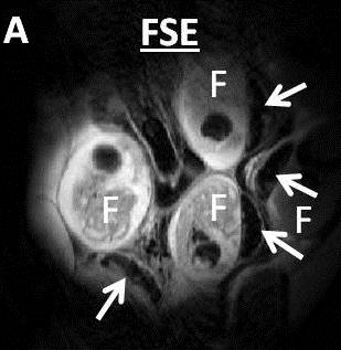
Anatomical image of a pregnant mouse showing multiple fetoplacental units.