Research Activities

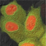
Cell culture showing cytoplasmic green and nuclear red fluorescence.

Protein separations at the nuclear envelope

The living cell separates its molecular components among numerous organelles. Most prominent is the nucleus, which encloses the chromatin and where synthesis of oligonucleic acids occurs. Communication between the nucleus and the cytoplasm takes place at the nuclear envelope, a double membraned structure perforated by large protein channels known as the nuclear pores. Through these channels the cell is able not only to pass but to concentrate specific proteins inside, or outside, of the nucleus. Biochemically the major players are a family of receptor proteins and a small GTPase, Ran.

Agrobacterium tumefaciens causes the common crown gall disease.

Agrobacterium tumefaciens and horizontal gene transfer

Nuclear uptake of foreign genes is the basis for many forms of viral infection, for gene therapy, and gene transfer without reproduction (horizontal gene transfer). Perhaps the most common case of horizontal gene transfer is the infection of plants by species of Agrobacterium. This common soil bacterium causes the crown gall disease, often seen for example on the trunks of trees. Having studied earlier the nuclear import of DNA by single molecule methods, we were motivated to examine this physiological example of DNA nuclear delivery. The mechanism is similar to bacterial conjugation, with a secreted protein, VirE2, that adapts to the eukaryotic host. We study the structure and interactions of VirE2 using a combination of biochemical, biophysical, and structural tools. Agrobacterium has also emerged as an important tool for genetic engineering of plants. We work together with the plant genetics lab of Prof Avraham Levy on site-specific targeting of DNA integration using Agrobacterium as a delivery vector.

Novel 3D imaging methods for cellular studies

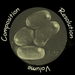
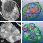
The lab is actively developing novel 3D modalities for ultrastructural imaging in cells. These include serial surface imaging by FIB-SEM and its correlation with light microscopy, soft X-ray cryo-tomography (SXT), and cryo-scanning transmission electron tomography (CSTET).

Synthetic protein bodies

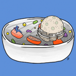
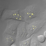
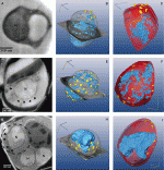
Organelles are often defined as membrane-bound compartments within the cell. Recent years have seen a conceptual expansion to include protein assemblies ranging from enzyme oligomers to nuclear bodies. The interactions governing the assembly are typically weak and may involve a collective phase transition from disperse to gel form. We explored the ability of the well-known ferritin protein to form large self-assemblies in tissue culture cells. The ferritin protein contains 4 alpha helices that self-assemble into a nearly spherical hollow cage, whose primary physiological function is in the storage of iron.

Plasmodium: the malaria-causing parasite

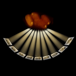
Malaria remains a widespread and devastating illness in many parts of the world. In collaborative projects we apply novel three-dimensional microscopy methods to study the blood-borne parasite that causes disease.

The Plasmodium parasite invade the red blood cells, where they multiply rapidly by schizogeny. Rather than dividing to pairs. Each cell multiplies into multiple daughters, typically 16, which acquire separate membranes only at the end of the process.