
TEDxWhiteCity - Shaping Life from the Shapeless: The Awesome Power of the Embryo
Dr. Karina Yaniv
Ministry of Science- נשים פורצות דרך במדע
![]()

|
In our lab we take advantage of the transparency and genetic amenability of the zebrafish embryo to uncover the mechanisms controlling specification of lymphatic endothelial cells and assembly of lymphatic vessels (Yaniv, 2006). Recently, we have revealed the existence of a novel niche of specialized progenitors, which gives rise to the lymphatic system, and have identified the first “lymphatic-inducing” signal. Moreover, using this factor we have been able to induce lymphatic differentiation of human embryonic stem cells, allowing for the first time the generation of human lymphatic endothelial cells in culture (Nicenboim, 2015).
Current projects in the lab involve further understanding of lymphatic formation in the zebrafish embryo, as well as characterization of organ-specific lymphatic vessels. In particular, we are interested in understanding how do lymphatic vessels in the heart form, what are their origins, and what is their putative role during cardiac pathologies. Finally, since the lymphatic system represents the main route for dissemination of metastatic cells, we aim to understand the mechanisms underlying tumor-induced lymphangiogenesis (the formation of new lymphatic vessels), and whether or not they relate to the circuits controlling lymphatic formation during embryonic development. |
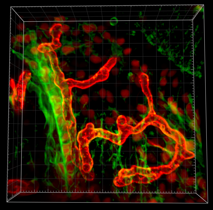
Specialized angioblasts in the floor of the Cardinal Vein give rise to the lymphatic system |
The formation of organ-specific vessels: At present it is well accepted that vessels of a particular organ display specific features that enable them to fulfill distinct functions. For instance ECs in the brain, which generate the blood-brain barrier, are structurally different from ECs in the fenestrated capillaries of the kidney or liver. We use transgenic zebrafish expressing fluorescent reporters in different organs, in combination with long-term live imaging to study the mechanisms underlying the formation of organ-specific vessels. In addition, we have developed protocols for UV-mediated photoconversion of restricted populations of ECs, followed by FACS isolation and RNA-Seq analyses. Altogether these studies are expected to shed light on novel players specifically expressed in ECs of certain organs, and responsible for their distinct function.

Lymphatic transdifferentiation and specialized vasculatureThe anal fin (AF), a bony locomotory organ that is established during metamorphosis. As it develops, it attracts rapid vascularization by secondary vessels—a specialized blood vessel type. Although extensive research has been devoted to understanding the physiology of this blood vessel type, its origins, molecular features and specialization mechanisms remained unclear. |
|
|
|
|
Neurovascular developmentNeurovascular development is the parallel emergence and patterning of the central nervous system (CNS) and the vascular system during embryogenesis. During this process the association and interaction of the different cells from the brain microenvironment (endothelial cells, pericytes, glia, neurons and microglia) results in an organized and functional structure known as neurovascular unit (NVU). Several studies conducted have demonstrated that these same cells are evolutionarily conserved in zebrafish. |
 |
|
|
|
Suppression of transcytosis activity across the blood brain barrier in zebrafishIn the project we are trying to find out what are the mechanisms for the suppressed transcytosis in the BBB using transgenic zebrafish lines which label specific cell types of the neurovascularunit such blood vessels, astrocytes, pericytes, neurons, glia and microglia |
 |