Putting the brakes on immune cells
How do circulating T cells come to a screeching halt at just the right moment?
Briefs
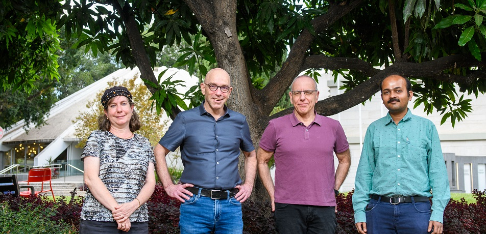
From left: Staff Scientist Dr. Sara Feigelson, Prof. Gilad Haran, Prof. Ronen Alon, and postdoctoral fellow Dr. Shirsendu Ghosh
Immune cells zip through our body’s blood vessels at dizzying speeds, braking at critical checkpoints with the precision of a plane landing on the flight deck of an aircraft carrier. This remarkable ability enables these cells to exit the vessels at just the right locations to cross into any site of injury or into lymph nodes, which they continuously patrol for signs of infection. But how do the cells detect these exit sites with such extraordinary precision and speed?
Weizmann researchers, led by Prof. Gilad Haran of the Department of Chemical and Biological Physics and rof. Ronen Alon of the Department of Immunology and Regenerative Biology, recently discovered that key immune cells called T lymphocytes keep the braking machinery right at their “fingertips.” These ingers are called microvilli and look like bumps on the surface of the T cells’ membrane. They help the cells sense and interact with their environment.
When a T lymphocyte cell needs to cross the wall of a blood vessel in order to get inside a lymph node, receptors on its microvilli “fingers” bind to specialized proteins on the vessel wall. Once fastened in this manner, the cell starts rolling along the vessel walls. As it does so, its receptors (known as chemokine receptors type7, or CCR7 in short) recognize small lymph node proteins called chemokines, which coat the inner surface of blood vessel walls.
The chemokines let the T cell know: You’ve arrived at the right spot in a lymph node—time to get off.
Surveillance system:
Only cells that have CCR7 molecules can read this exquisitely targeted message. The CCR7 transmit the signal further, activating an adhesion protein called LFA-1 integrin, which in turn binds to sticky molecules on the blood vessel wall. The T cell can then come to a sudden halt, exit the vessel, and enter the lymph node—where it spends anywhere from a few minutes to several hours on the lookout for foreign particles, before returning to the blood circulation if it doesn’t encounter anything troublesome. Thousands of such events take place within hundreds of lymph nodes around our body every minute, ensuring effective immune surveillance.
In earlier research, Prof. Alon and his team found that the T cells’ braking signal is generated in under half a second. To learn how the cells manage to put together their braking machinery so quickly, Prof. Alon teamed up with Prof. Haran, whose lab has developed a new super-resolution microscopy method for exploring cell surfaces at the level of single molecules.
One important conclusion they found from their collaborative study: the machinery the T cells need for their rapid braking is pre-assembled on the tips of their microvilli. “This explains how they manage to transmit the braking signal within less than half a second,” Prof. Alon says.
These findings not only shed new light on how T cells perform immune surveillance of the body, but also open up a new line of research into mechanisms guiding the movement of other cell types, including tumor cells—which, while flowing through blood vessels, come to a halt at sites of metastasis.
Prof. Ronen Alon is supported by:
- Moross Integrated Cancer Center
- Prof. Alon is the incumbent of the Linda Jacobs Chair in Immune and Stem Cell Research
Prof. Gilad Haran is supported by:
- Deloro Institute for Advanced Research in Space and Optics
- Nancy and Stephen Grand Center for Sensors and Security
- Henry Chanoch Krenter Institute for Biomedical Imaging and Genomics
- Prof. Haran is the incumbent of the Hilda Pomeraniec Memorial Professorial Chair