

The high percent of point defects in oxygen deficient ceramics is central to many important macroscopic properties displayed by these materials: e.g. Ionic conductivity, catalytic properties, Electro mechanic, and time dependent elastic properties (Anelasticity). Our group is working on answering several fundamental scientific questions related to defect sites. For example: What is the arrangement of atoms around defect sites? How does defect concentration or defect identity affect the local structure? Can we correlate microscopic structural changes to macroscopic properties such as electro-mechanical activity? How do defects affect the vibrational dynamics (phonons) of these highly defective materials?
X-ray absorption spectroscopy
A powerful tool we are employing in order to study the microscopic structure of ceramics is X-ray absorption spectroscopy. This technique allows us to access the local inter-atomic environment of point defects and even access changes in local structure in-situ (under applied electric field). X-ray absorption spectroscopy requires a high energy X-ray photon source (synchrotron) and we have therefore held a long standing collaboration with the group of Prof. Anatoly Frenkel at Stoney Brook University and at the Brookhaven national lab in NY, USA.
Frenkel group website: https://www.bnl.gov/chemistry/SDAN/index.php
Schematic of the experimental set up for acquisition of X-ray absorption in the high-energy resolution fluorescence detection (HERFD) configuration from a doped ceria thin film system, under application of electric field (in-situ). The layers of the sample are detailed on the right hand side. Image adapted from: Li Y. et al.(2016). Geometry of electromechanically active structures in Gadolinium - doped Cerium oxides. AIP Advances. 6:(5) 055320. 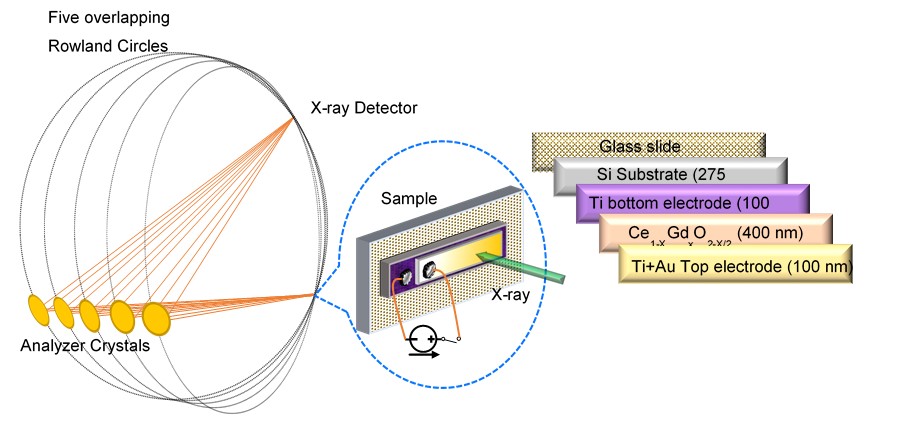
Key publications
Kraynis O., Timoshenko J., Huang J., Singh H., Wachtel E., Frenkel A. I. & Lubomirsky I.(2019). Modeling Strain Distribution at the Atomic Level in Doped Ceria Films with Extended X-ray Absorption Fine Structure Spectroscopy. Inorganic Chemistry. 58:(11)7527-7536.
Li Y., Kraynis O., Kas J., Weng T., Sokaras D., Zacharowicz R., Lubomirsky I. & Frenkel A. I.(2016). Geometry of electromechanically active structures in Gadolinium - doped Cerium oxides. AIP Advances. 6:(5)
Korobko R., Lerner A., Li Y., Wachtel E., Frenkel A. I. & Lubomirsky I. (2015). In-situ extended X-ray absorption fine structure study of electrostriction in Gd doped ceria. Applied Physics Letters. 106:(4)
Raman spectroscopy
Raman spectroscopy can sample the phonon modes (lattice collective vibrations) of a crystalline material. In defect rich ceramics, one can observe modes emerging from the crystal average symmetry (space group), as well as new modes emerging from the presence of point defects which locally distort the average symmetry. In addition defects can affect the behavior of the main (average) phonon modes. For example, we have shown that the sensitivity of the main symmetric vibration mode in doped ceria to anisotropic strain, is reduced in thin film structures. We have also observed time dependence of the mode energy in thin films (indicative of anelasticity). We continue to study fundamental aspects of the Raman spectra of oxygen deficient materials and their connection to defects and mechanic properties.
A. Schematic of a substrate supported doped ceria film undergoing silicon substrate removal to form a self-supported membrane (membrane thickness ~1micron). B. The Raman spectrum of the ceria film, before (dashed, black) and after (continuous, green) substrate removal. The observed shift to lower energies (Red shift) in the main Raman mode (depicted in the inset) is due to change in compressive strain magnitude between the substrate supported and self-supported films. The Frontispiece cover image on the right hand side is an artistic interpretation of sampling the ceria crystal structure with the red He-Ne laser during Raman acquisition: on a membrane surface (Front unit cell) and on a substrate supported surface (background unit cell). 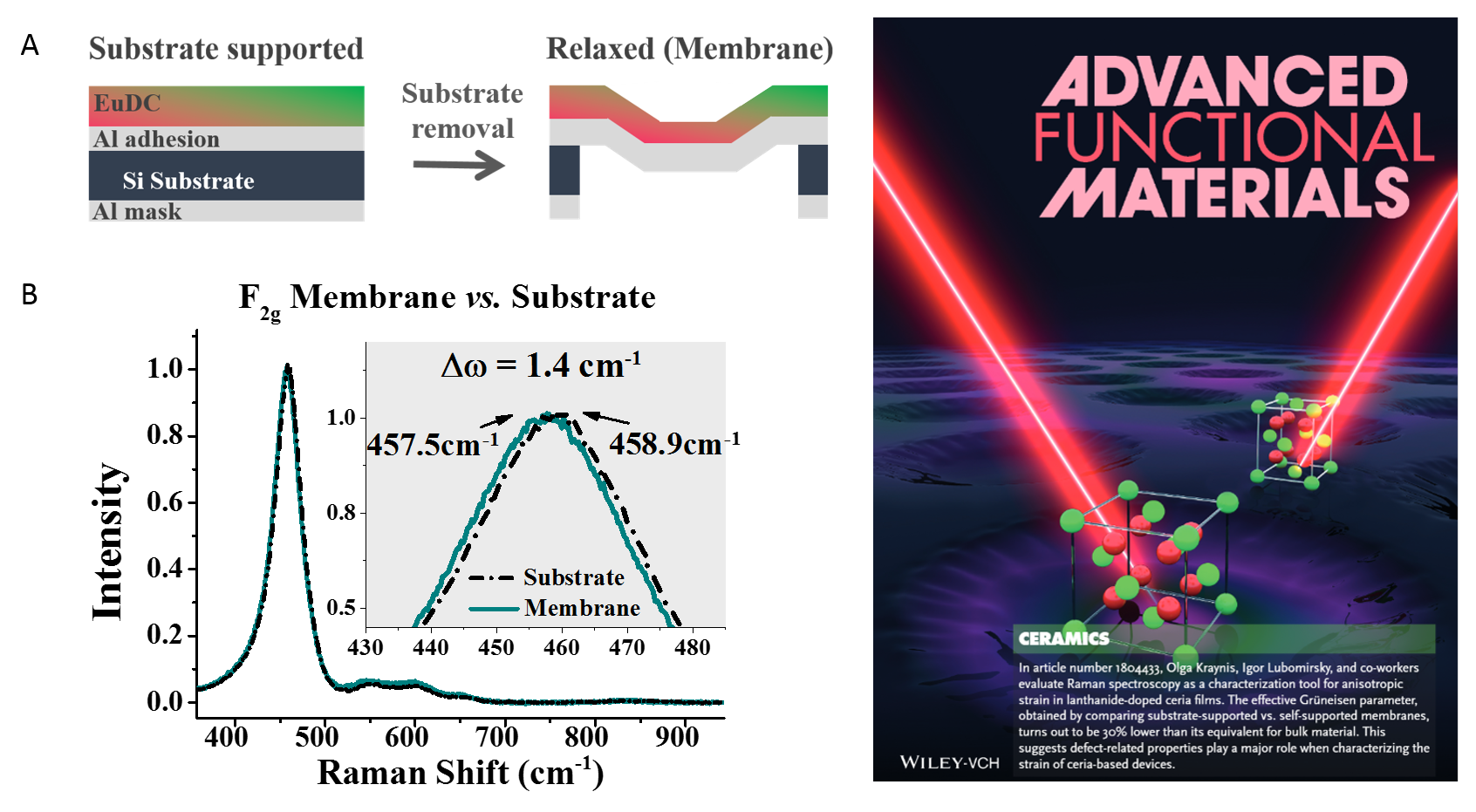
Key publications
Kraynis O., Makagon E., Mishuk E., Hartstein M., Wachtel E., Lubomirsky I. & Livneh T. (2019).Suitability of Raman Spectroscopy for Assessing Anisotropic Strain in Thin Films of Doped Ceria. Advanced Functional Materials. 29:(11)
Kraynis O., Wachtel E., Lubomirsky I. & Livneh T. (2017). Inelastic relaxation in Gd-doped ceria films: Micro-Raman spectroscopy. Scripta Materialia. 137:123-126.