Skip to main content
Skip to navigation
Accessibility
Increase font size
Decrease font size
Sharpen color
Grayscale
Invert color
Default
Disclaimer
close
Toggle navigation
Menu
Department of Molecular Genetics
Karzbrun Lab
Self-Organization in Embryonic Development
Home
You are here
Home
›
Research
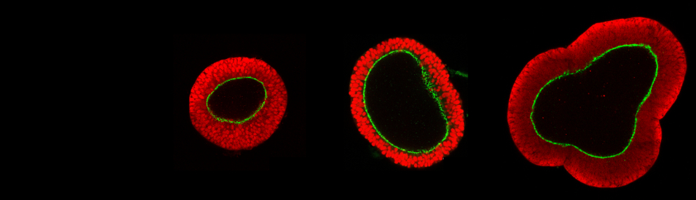
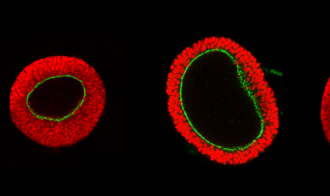
Research