

Using reversal in DSGCs to shed new light on the mechanisms underlying direction selectivity
Direction selective retinal ganglion cells respond strongly to an image moving in one direction (the “preferred” direction) and weakly to an image moving in the opposite, “null”, direction. The asymmetric computation is thought to be hardwired, arising from asymmetric inhibitory inputs from starburst amacrine cells (SACs). Yet, we have discovered that the directional preference of a neuron can be altered and reversed by 180 degrees following a short repetitive visual stimulation. This reversal is robust and long lasting, and occurs not only in juvenile but also in adult mice.
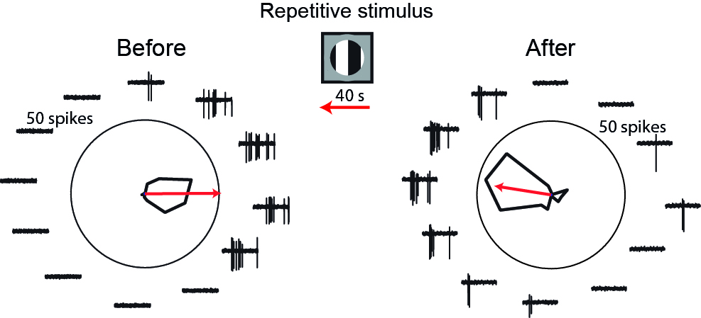
Direction selective ganglion cells (DSGCs) reverse their directional preference
Directional tuning of a DSGC before (left) and after (right) repetitive stimulation with 40 sec of drifting grating (shown in center), revealing reversal of directional preference. Polar plot represents number of spikes in response to 3 sec gratings drifting in 12 directions. Red arrow indicates the preferred direction. Traces show examples of 0.5 sec activity.
Adopted from Rivlin-Etzion et al., Neuron 2012
To understand how DSGCs overcome the circuit’s anatomy and reverse their directional preference, we record from neurons in the direction selective circuit using different patch-clamp techniques and in response to various moving stimuli. A major focus is the SAC that is known to mediate the directional response in DSGC. Our data sheds new light on the multiple mechanisms that underlie direction selectivity and how they can act in orchestra or oppose one another.