Research

Follicular regulation of meiotic resumption and oocyte maturation inhibitor

spontaneous maturation of isolated cumulus-enclosed oocytes (CEO) in culture led Pincus and Enzmann (1935) to suggest that follicle cells somehow inhibit the resumption of meiosis. We have tested this assumption by co-culture of porcine cumulus-enclosed oocytes (CEO) with other follicular constituents, such as porcine follicular fluid (PFFI), granulosa cells or ovarian theca-somatic tissue.

The ovulatory response

Culture of pre-ovulatory follicles provided insights far beyond the initial aim to examine the regulation of resumption of meiosis. The ability of gonadotropins to induce oocyte maturation in explanted follicles, without causing follicle rupture or ovulation, as well as the ability to inhibit follicular luteinization without affecting resumption of meiosis, led to the understanding that these three biological responses of ovulation, all initiated by the gonadotropic surge, are separable and may involve distinct and independent mediatory pathways downstream to gonadotropin binding.

Atresia as an Apoptotic Process

The vast majority of the growing follicles undergoes degeneration, through atresia, rather than ovulation. Examination of the mechanisms which determine whether a follicle will ovulate or undergo atresia was impeded due to the ability to recognize atresia only in retrospect.

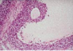
In vitro model for studying the regulation of oocyte maturation

Oocyte meiosis in mammals is a protracted process, subject to several stop-go controls. The oocyte initiates the first meiotic division during embryonic life or shortly after birth. Meiosis is arrested, in most mammals, in neonatal life at diplotene/dictyate stage of the first meiotic prophase. The hallmark of the dictyate oocyte is the large nucleus known as “germinal vesicle” (GV).