
Recent Studies
Bone 3D Structure and Function (together with Ron Shahar)
A key discovery made in 1986 by Weiner and Traub (Weiner and Traub, 1986) was that the individual mineralized collagen fibril, the basic building block of bone) does not have radial symmetry, but its structure is in essence crystalline. This is due to the arrangement of the plate-shaped crystals in the form of layers across the entire fibril. This discovery necessitated the reanalysis of structure of the most common materials composed of mineralized collagen fibrils: lamellar and other bone types, dentin and tendon. This resulted in the proposal of a model for lamellar bone in which the orientations of the crystal arrays change from one side of an individual lamella to the other (Weiner et al., 1997). Recently we have also investigated the 3D organization of the collagen fibrils in lamellar bone using a dual beam electron microscope and the serial surface method. Reznikov et al discovered in this way that in rat lamellar bone, the major components are indeed aligned in differently oriented arrays of mineralized collagen fibrils, but surprisingly there is also a relatively disordered layer in each lamella (Reznikov et al., 2012). Feingold et al showed in horse bone that this disordered component is even more prominent than in rats (Faingold et al., 2013). Reznikov et al also showed that one component of the canalicular network is closely associated with this disordered layer. The latter also has less mineral than the other layers within a lamella, and might well undergo much more strain when the bone is mechanically loaded. We thus postulate that this disordered layer might have a critical function in bone mechanics and sensing (Reznikov et al., 2012).
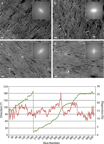 |
Fig. 4. Four images from the 3D stack 6.3 μm thick (specimen #6); (a) slice #320, (b) slice #390, (c) slice #450, (d) slice #600 and (e) a computer generated texture analysis of the collagen fibrils based on the FFT of each image in the stack. The texture analysis shows the preferred fibril direction (green), and fibril dispersion (red). The bone long axis direction is oriented at 90° (vertically). Panel a shows oblique alignment of the fibrils and moderate angular dispersion. Panel b shows “disordered” fibril organization. Panel c shows oblique alignment of the fibrils and low angular dispersion. Panel d shows horizontal alignment of the fibrils and low angular dispersion. The insets show the frequency domains (FFT) of each image. The canaliculi cross-sections are spindle-shaped in panels a, c and d and their long axes are aligned with the preferred orientation of the fibrils. In panel b the canaliculi cross-sections are roughly circular and the FFT shows almost no preferred direction of the fibrils. In panel e, note that the break at slice 180 is an artifact as 180° continues at 0°. Scale bars 400 nm. (From Reznikov et al, 2012) |