
Trabecular bone is an intricate 3D network of struts and plates. We developed a parameter that measured the angle between two connected trabeculae – the Inter-Trabecular Angle (ITA). An analysis of the inter-trabecular angles (ITAs) of the 3D fabric of the trabecular bone of the human proximal femur reveals the presence of highly conserved underlying topological motifs that include most significantly nodes with 3, 4 and 5 emanating edges in proportions of 12:4:1.
Recent Studies
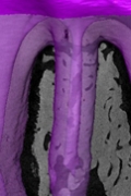
The prevailing question we address is how does the whole tooth-periodontal ligament-alveolar bone complex function during mastication? We use a top down approach in this research, and have taken advantage of optical metrology methods that make it possible to map deformations on the surface of a tooth (Lev-Tov Chattah et al., 2009; Zaslansky et al., 2006a; Zaslansky et al., 2006b). We have shown that when loaded both the tooth itself deforms, as well as the soft periodontal ligament (PDL) in ways that complement each other.

A key discovery made in 1986 by Weiner and Traub (Weiner and Traub, 1986) was that the individual mineralized collagen fibril, the basic building block of bone) does not have radial symmetry, but its structure is in essence crystalline. This is due to the arrangement of the plate-shaped crystals in the form of layers across the entire fibril. This discovery necessitated the reanalysis of structure of the most common materials composed of mineralized collagen fibrils: lamellar and other bone types, dentin and tendon.

Ever since the study of the lens arrays in the brittlestar that started in our group (Aizenberg et al., 2001), we have been fascinated by the possibility that light manipulation by biogenic minerals is a widespread phenomenon, not only to produce structural colors, but also possibly on a huge scale in the plant kingdom that after all, “makes its living” by manipulating light. We have elucidated the structural basis by which fish and spiders produce metallic lusters based on ordered stacks of extremely thin arrays of guanine crystals (Levy-Lior et al., 2010).

In the almost 25 years since the late Heinz Lowenstam and I published the book entitled “On Biomineralization” (Lowenstam and Weiner, 1989) some fundamental concepts still prevail (eg the preformed extracellular matrix into which mineral forms), but some basic paradigms have changed. The concept that the first minerals are often deposited in vesicles within cells and are then transported into the extracellular space where they often undergo a transformation into more ordered forms, is rapidly becoming established (Fig. 1).