A STEM view of chromatin
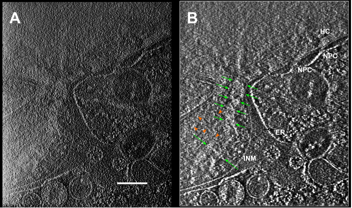
Cryo-STEM tomography of fibroblast cells near the nuclear envelope revealed unexpected large-scale structures extending through the nuclear volume. These appear both in regions associated with heterochromatin and euchromatin. A close look with stereo pair imaging reveals a spiral cylinder or "slinky" shape, with a diameter of about 200 nm. We propose that the nucleosome chain may form such a slinky naturally, and that its expansion or contraction may govern access to the chromatin. Notably, the 3D imaging was performed on a cultured cell in its native state, fully hydrated without chemical fixation, embedding, staining, or sectioning of any sort. 3D deconvolution processing made a crucial contribution to noise reduction and to axial resolution. This work was a collaboration with Prof John Sedat of UC San Francisco.