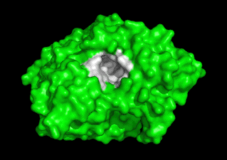
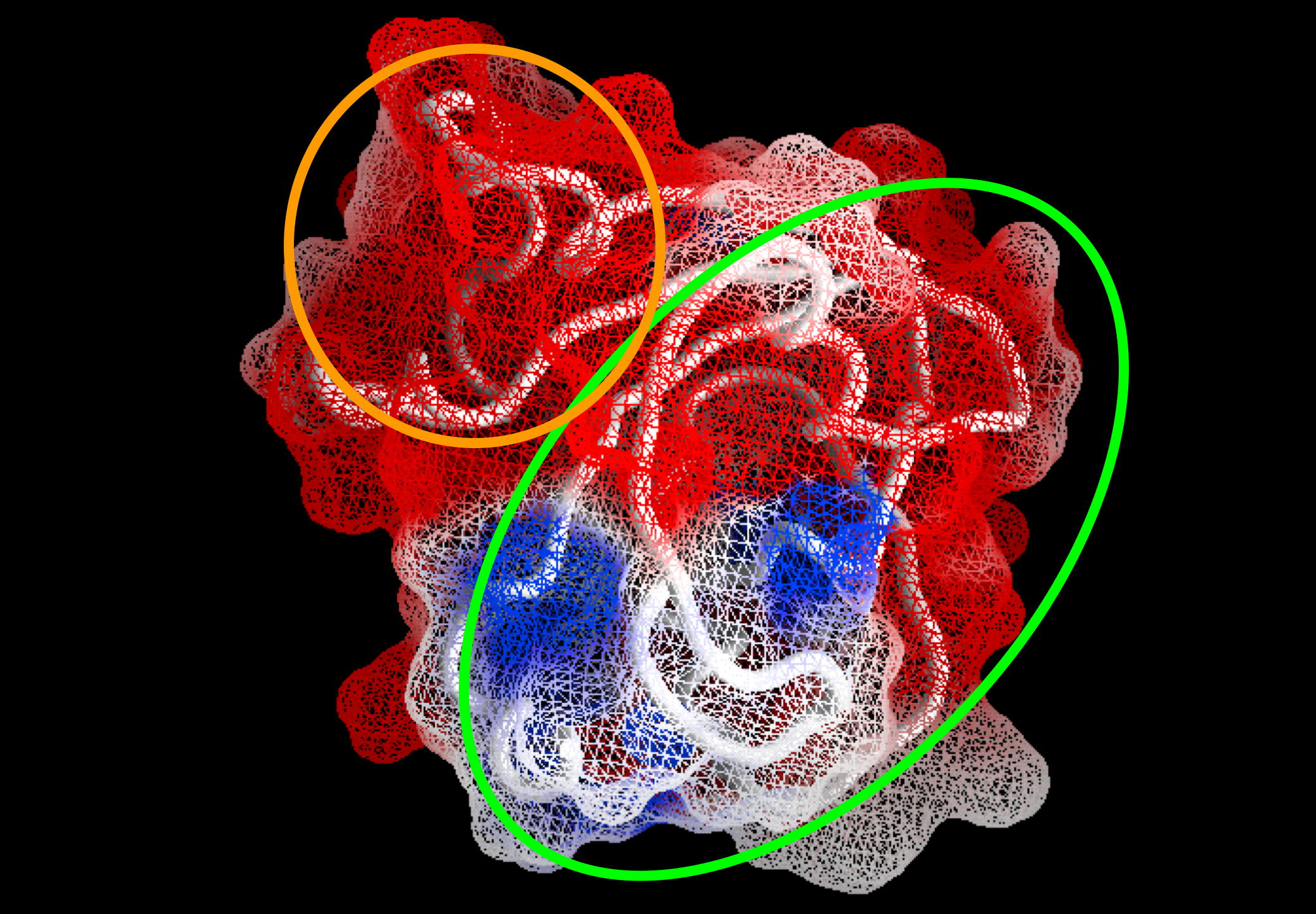
Other Projects


Structure of the binding site of the AChR as visualized in the X-ray structure of a complex between α-bungarotoxin and a high affinity mimotope peptide

Together with Sarah Fuchs and Ephraim Katzir, we have determined the crystal structure of a complex of α-bungarotoxin with a high affinity 13-residue peptide homologous to the binding region of the α-subunit of the AChR.


The peptide fits snugly to the toxin and adopts a β hairpin conformation. The structures of the bound peptide and the homologous loop of ACh-binding protein, a soluble analogue of the extracellular domain of AChR, are remarkably similar. Their superposition suggests that the toxin wraps around the receptor binding-site loop and, in addition, binds tightly at the interface of two of the receptor subunits, where it inserts a finger into the ligand-binding site, thereby blocking access to the ACh-binding site, thus explaining its strong antagonistic activity .

AChR binding
Fig. 1. The combined model of a-BTX-HAP (red) and AChBP structure with subunit A in green and subunit B in yellow, showing the insertion of loop 2 of the toxin into the interface of the two subunits. The positively charged HEPES molecule (black stick figure) shows the location of the acetylcholine-binding site and the blockage of passage to this site caused by the binding of the toxin. The HAP, which overlaps the 182-193 loop of AChBP, is shown in blue.


WAT/PRAD Complex, a model of the tetramerization domain of AChE

The 'tailed' heteromeric molecules are the most physiologically important forms of AChE, and the predominant forms in brain and at neuromuscular junctions. Massoulie and colleagues pinpointed a small proline-rich attachment domain (PRAD), around which the globular subunits assemble to form tetramers. The critical feature of this 17-residue peptide is the presence of three and five consecutive prolines; thus even synthetic polyproline can replace the natural PRAD.

Similarly, a well-conserved 40-residue peptide at the C-terminus of the AChE subunit sufficies to form the same quarternary organization by interacting with PRAD. This portion of AChE contains a series of seven aromatic residues, including three equally spaced Trp residues. Thus, it was named the 'tryptophan amphiphilic tetratmerization' (WAT) domain. We have crystallized a complex of this WAT/PRAD complex (ratio 4:1), with selenomethionine incorporated, and plan to determine its structure using MAD.


Radiation damage is an inherent problem in X-ray crystallography

It has commonly been assumed to be non-specific, and manifested as gradual decay in the data quality as data collection proceeds, even at cryo temperatures. Recently, in preparation for time-resolved crystallography experiments, we collected nine successive complete data sets on the same TcAChE crystal, at 100K, on a powerful undulator beamline at the ESRF. We found that radiation damage can be highly specific. Thus one disulfide was cleaved completely, as in Fig. 2 below, while another was resistant. Highly exposed COOH groups, and those in the active site, are especially susceptible, and active-site H440 much more than others. Our findings have practical implications for routine data collection at high-energy synchrotrons and for interpretation of data so obtained. They also offer a direct approach to studying the radiation chemistry of proteins and nucleic acids, and may yield information concerning flexible regions or putative weak links in a given macromolecule.

Radiation Damage
Fig. 2. X-ray radiation from a synchrotron source can rapidly cause specific damage to a protein, as seen by X-ray crystallography. The four images represent a series of electron density maps collected from a single AChE crystal. Note the disappearance of the disulfide bond.

movie icon View our group's movies about this topic: