Making cancer history
News from the Moross Integrated Cancer Center
Features
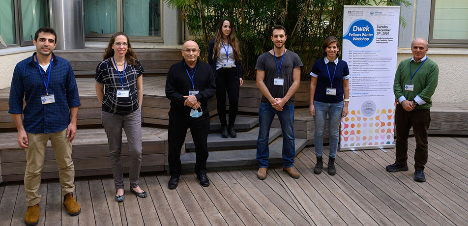
L to R: Omer Goldman, Hila Tishler, Prof. Avigdor Scherz, Hadas Lewinsky, Akhiad Bercovich, Reut Riff, Prof. Yossi Yarden
It was back in 1775 when English surgeon Percivall Pott, reporting on the prevalence of carcinoma among London chimney sweeps, identified the first clear link between cancer and the environment.
A century later, Brazilian ophthalmologist Hilário de Gouvêa proved that cancer susceptibility is genetic — that is, it can be passed down from a parent to a child.
Today, these fundamental realizations form the basis of investigations conducted under the auspices of the Moross Integrated Cancer Center, the Weizmann Institute’s flagship framework for cancer research, housed in the Ullman Building of Life Sciences. At the same time, Moross Center scientists are contributing paradigm-shifting discoveries that illuminate entirely new paths toward better cancer prediction, prevention, diagnosis, and treatment. Among discoveries achieved by dozens of Moross-associated labs over the past year, the following studies illustrate the Center’s impact, which is bringing us steadily closer to the ultimate goal: the defeat of cancer in all its forms.
A bacterial ID card for tumors
Several years ago, Dr. Ravid Straussman, a member of the Department of Molecular Cell Biology, discovered bacteria lurking within tumor cells of the human pancreas. These bacteria were shown to “digest” and inactivate chemotherapy drugs, thereby blocking the drugs’ anti-cancer effects. Now, based on a study of over 1,000 tumor samples of different human cancers, Dr. Straussman has shown that unique populations of bacteria reside within each tumor type.
This research, performed together with Dr. Noam Shental from the Department of Mathematics and Computer Science at the Open University of Israel and published in the journal Science last year, suggests that characterizing interactions between cancer cells and their resident bacteria may lead to new strategies for improving personalized cancer treatment. In the future, it may also become possible to manipulate intra-tumor bacterial populations in order to make treatment more effective.
“Most baceria you find in tumors are known to be present normally in people, but there’s also a minority of bacteria we found that were never described in humans or any other host before,” Dr. Straussman says. “We also found bacterial populations inside the immune cells that reside within cancerous tumors—a discovery that has implications for cancer immunotherapy.”
The Straussman team surveyed seven different cancer types: breast, lung, ovarian, pancreatic, melanoma, bone, and brain. Examination of these samples revealed that about 70 percent of breast cancer tumors are characterized by the presence of bacteria.
Moreover, the scientists discovered that the complement of bacterial populations found in breast tumors is particularly complex. In another intriguing finding, revealed when the scientists compared lung tumors from smokers to tumors from patients who never smoked, Dr. Straussman discovered that bacteria from smokers’ lung cancer cells had a qualitative difference: these bacteria had many more genes for metabolizing nicotine, toluene, phenol, and other cigarette-related chemicals.
By shedding light on how the balance of bacterial populations in cancerous tumors influences the dynamics of tumor ecology, this research may “reframe” the protocols used to diagnose and treat human cancers, something that may eventually lead to more hopeful cancer prognoses.
Finding the red flags
Prof. Yardena Samuels, also from the Department of Molecular Cell Biology, discovered a gene that is mutated in 32 percent of colorectal cancer patients as well as many other human cancers. She uses DNA sequencing to identify groups of genetic mutations involved in melanoma, and to differentiate these from “passengers”—genetic abnormalities that play no role in tumorigenesis.
In a recent achievement, Prof. Samuels reported on a new method that may make it possible to create improved and highly personalized treatments for patients with melanoma. She has identified “red flags”—mutated peptides that appear on melanoma cells’ outer membranes, which allows the immune system to identify these cancer cells as foreign, and target them for attack. Prof. Samuels’ findings offer a powerful alternative to current methods that seek to activate immune T cells against these mutated surface peptides, which are known as neo-antigens.
One of the problems in uncovering neo-antigens in cancers like melanoma, however, is that they are presented by a protein complex called HLA–a complex that can come in thousands of versions, even without the addition of cancerous mutations. Indeed, the algorithms often used to search the cancer-cell genome for possible neo-antigens have predicted hundreds of candidates. This makes it extremely challenging for pharmaceutical companies to use neo-antigen targeting as the basis of a “one-size-fits-all” immunotherapy drug.
Now, Prof. Samuels and her colleagues have devised a new way to identify neo-antigens of melanoma cells, together with the specific T cells “primed” to respond to them. The group’s strategies were so robust that their neo-antigen-specific T cells killed 90 percent of the target melanoma cells both in laboratory plates and in mice.
She then went on to demonstrate that cancer cells from different metastases within the same patient have neo-antigen “profiles” that are remarkably uniform. This discovery may eventually contribute to clinical strategies in which doctors would create personalized drug regimens that would stimulate T cells to destroy not just an individual patient’s primary tumor, but secondary malignancies as well.
A simple blood test for lung cancer risk
Most cases of lung cancer—the leading cause of cancer death—are linked to smoking. That puts smokers first in line for medical screening procedures that seek to identify lung tumors early, so that patients can begin treatment and improve their chances for long-term survival.
Today, screening protocols are traditionally based on age and smoking status. However, reliance on these two risk factors alone misses the mark, as preventative screening in this selected population fails to identify most lung tumors. Moreover, individuals who are not obvious candidates for screening remain unaware of the danger they may face – something that leads to delayed treatment and poorer prognosis.
Prof. Zvi Livneh and senior staff scientist Dr. Tamar Paz-Elizur, both members of the Department of Biomolecular Sciences, in collaboration with researchers from the UK, have proposed a new lung cancer screening protocol based on a simple blood test. Relying on individual patients’ “DNA repair scores”—a summation of the activity of three DNA repair enzymes through which cells are known to respond to genetic damage—the new screening strategy accurately identifies individuals at heightened risk for lung cancer.
Based on the examination of hundreds of British patients, the study demonstrated that a low DNA repair score reveals a five-fold greater risk for lung cancer onset than would typically be estimated based on age and smoking status alone. A low DNA repair score may also help explain why some non-smokers (normally not referred for preventative screening) develop lung cancer, thereby contributing to the development of clinical criteria for promoting early diagnosis in the non-smoking population. These findings have important implications for improving the effectiveness of lung cancer screening, and providing more at-risk patients with access to early diagnosis and treatment.
In another, unexpected finding that emerged from this study, Prof. Livneh’s team found that a low DNA repair score in lung cancer patients, but not in healthy people, correlates with a broad increase in gene expression pathways that mediate the body’s immune response. This indicates that DNA repair score data—as revealed in a blood test—could potentially contribute to personalized treatment, by helping doctors predict how individual lung cancer patients will respond to cancer immunotherapy.
Saluting the Dwek Fellows
young leaders in cancer science
The Dwek Institute for Cancer Therapy Research, one of the three pillars of scientific investigation operating under the auspices of the Moross Integrated Cancer Center, is dedicated to supporting activities that contribute to clinicians’ ability to treat and defeat cancer. Recently, the Dwek Institute launched a new initiative: the Dwek Fellowship Program, which recognizes and supports outstanding PhD students and postdoctoral fellows who are actively pursuing studies aimed at developing novel anti-cancer strategies.
Supervised by Prof. Yosef Yarden, Director of the Dwek Institute for Cancer Therapy Research, the selection of the inaugural cadre of Dwek Fellows was a two-tier process. First, a call went out to Weizmann life science laboratories involved in oncology-related research, requesting nominations for up to two PhD or postdoctoral candidates. This resulted in 20 applications, from which an academic committee selected ten finalists. In December, the 10 finalists were invited to present their research at a small workshop.
The event kicked off with remarks by Prof. Avigdor Scherz, a member of the Department of Plant and Environmental Sciences who is co-inventor of TOOKAD-VTP ®, a photodynamic treatment that cures early-stage prostate cancer and is being developed for use in other cancer types. The fellowship finalists offered some inspiring words of their own, describing how, through their individual research, they hope to contribute to improving cancer treatment.
At the conclusion of the program, the finalists themselves ranked the presentations, with the five highest-ranked finalists named winners of the Dwek Fellowship.
Here’s a look at the first cadre of Dwek Fellows
Hila Tishler is pursuing her PhD in the lab of Prof. Ayelet Erez in the Department of Biological Regulation. After a two-year stint at a biotech company where she helped develop a cancer immunotherapy drug, she began doctoral research at the Weizmann Institute, where she focuses on the metabolic cross-talk between cancer cells and their microenvironment. Her recent experiments have shown how, by altering the availability of an amino acid that mediates the metabolism of nitrogen, it is possible to reduce tumor growth, affect T cells’ anti-cancer activity, and potentially sensitize cancer cells to the body’s immune defenses—which could potentially improve patients’ response to immunotherapy.
Dr. Reut Riff is a postdoctoral fellow in Dr. Ravid Straussman’s lab. She focuses on bacteria-mediated anti-cancer vaccines, an approach that seeks to optimize cancer patients’ response to immunotherapy by exploiting bacterial activation of the body’s immune response. Building on the recent discovery in the Straussman lab that specific bacterial populations reside in particular tumor types, Dr. Riff is trying to establish a pipeline for identifying bacteria that will home to a tumor, and manipulate it to express tumor-specific neo-antigens. This would make it possible to use such bacteria to activate the targeting of T cells, thereby making cancer immunotherapy more effective.
Hadas Lewinsky is pursuing her doctorate under the supervision of Prof. Idit Shachar in the Department of Immunology. She is focused on “suppressors”—cells in the tumor microenvironment that help shelter cancer cells from attack. Research in the Shachar lab has shown that a receptor called CD84 is highly expressed in suppressor cells in the microenvironment of multiple myeloma. It has also shown that blocking this receptor leads to a more effective anti-cancer immune response. Hadas is conducting experiments in solid tumors showing similarly positive results.
Omer Goldman is pursuing his doctorate in cancer metabolism in the lab of Prof. Ayelet Erez. He is focused on the urea cycle, a mammalian pathway localized in the liver that prevents the accumulation of toxic levels of nitrogen in the body. Experiments show that, early in tumor development, significant “rewiring” is seen in liver metabolism, as well as in compwonents of the immune system—even in cancers that do not metastasize to the liver. Preliminary studies in cancer patients provide evidence of similar metabolic dynamics, suggesting that therapeutically blocking this metabolic rewiring in the liver could potentially provide clinical benefits.
Akhiad Bercovich is a PhD candidate in the laboratory of Prof. Amos Tanay in the Department of Computer Science and Applied Mathematics. Akhiad is developing computational models of gene expression data captured using single-cell genomics approaches. Such methods make it possible to identify and describe conserved behaviors of immune cells from the tumor microenvironment, in both mouse cancer models and human patients. Akhiad is currently developing tools to clarify cellular responses to immunotherapy drugs in many cancer types, while using his model systems to predict and experimentally validate novel combination treatment regimes.
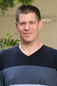
Dr. Ravid Straussman
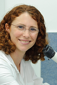
Prof. Yardena Samuels
MICC Building