New materials, inspired by life
The science of biomimcry and bioinspiration at the Center for Advanced and Intelligent Materials
Features

Yellow tube sponges from the ocean floor
Part organic, part inorganic: researchers at the Weizmann Institute of Science are merging the best of both worlds. In laboratories on campus, the innate qualities of biological materials—optimized over 500 million years of evolution—are increasingly being engineered into new, hybrid materials and technological approaches. From bio-molecular sensors to synthetic gene circuits, to hybrid materials with qualities that match or exceed those found in nature, scientists are harnessing the patterns and principles behind the dynamic processes of life. This represents a major avenue of exploration in the Center for Advanced and Intelligent Materials, a new Institute flagship project.
The organic-inorganic “fusion” has become more prominent in the past few years, fueled by improved imaging technologies that allow scientists to analyze the structure of living things on the molecular and even the atomic level. Nanotechnology also plays a role, providing new tools that make it possible to fabricate hybrid materials that combine non-biological elements with DNA and proteins. And the specificity with which organic molecules bind to targets renders them excellent “nano-tools” for achieving very specific biotechnological results.
Integrating materials science, biomedical engineering and computational biology, and emerging approaches from the worlds of biology, chemistry, and physics, researchers are co-opting nature’s hidden talents in order to drive progress at the forefront of nanotech innovation.
A speedy “snapshot”
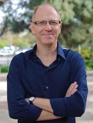
To use biological materials, scientists first need to understand how they work. This is the focus of Prof. Gilad Haran, who has devised a light-based platform for probing the dynamic—and incredibly speedy—structural changes that are critical to the function of individual molecules.
“Biomolecules, specifically proteins, operate like tiny machines. We are interested in the dynamic process by which these nano-machines adopt various conformational shapes, corresponding to different functions,” says Prof. Haran, a member of the Department of Chemical Physics. He has developed a tool that allows his team to observe and measure motion in an individual biomolecule as it occurs over windows of time lasting just one millionth of a second.
Prof. Haran’s system starts with nanotechnology. He equips individual proteins with a pair of light-sensitive antennae: a “donor” that emits green photons when excited by laser light, and an “acceptor” which, after receiving energy from the donor, emits photons that are red. This system makes it possible to collect and measure the light emitted at these two different wavelengths. Trapping these antennae-equipped proteins inside lipid vesicles tethered to a surface makes it possible to monitor the proteins’ light-based signals over time. The result, says Prof. Haran: “We can ‘see’ the physical distance between the two antennae, and also track dynamic changes in molecular structure. It gives us the power to ‘spy’ on an individual, biological molecule in action.”
Using this system, Prof. Haran revealed how, on the road to a particular conformation, proteins “rest” in a series of intermediate folding states. He also demonstrated that such conformational changes can take place at an astounding speed: in a study of an enzyme involved in the regulation of ATP—the energy “currency” of biological molecules—his technique showed that this protein opens and closes 100,000 times per second.
“The more we understand about protein dynamics, the better we will be able to mimic these properties in synthesized, biotechnological systems,” he says.
“Sniffing out” cellular characteristics—the molecular nose
Another Weizmann researcher working with individual molecules is Dr. David Margulies of the Department of Organic Chemistry, who has pioneered a method for synthesizing “nosy” molecules capable of peering into cells and see how their work is done.
In traditional biochemistry, when scientists want to investigate what’s going on inside a cell, they often use probes—antibodies or other molecules—that can bind one specific protein. When successful, this protein hunt results in a fluorescent signal that can be read by an optical microscope. But it takes multiple experiments to reveal the many factors comprising a cell’s chemical profile.
A “molecular nose” invented by Dr. Margulies offers a new solution: It generates quick and highly detailed analytic results, revealing and quantifying the presence of a number of proteins in a single experiment. Why does he call it a nose? Because, just as a small number of olfactory receptors allow the human nose to differentiate between vast numbers of smells, a small number of molecular receptors allow Dr. Margulies’ molecule-size device to differentiate between multiple proteins, sensing and quantifying them within living cells. The system reveals the relative balance between the proteins it senses by generating an optical signature—a multi-colored, fluorescent “report” of a living cell’s unique chemical identity. Successfully applied in scenarios requiring simultaneous sensing of multiple factors, the molecular nose has been used to characterize the chemical content of the non-regulated medications that have been associated with drug counterfeiting in developing countries.
The sensor’s potential was also demonstrated in a recent study published by Dr. Margulies in the Journal of the American Chemical Society. Here, the molecular sensor successfully differentiated between, and quantified, various states of amyloid beta aggregation—the progressive “clumping” of mis-folded proteins that is the hallmark of Alzheimer’s disease.
“In order to better understand the factors that influence Alzheimer’s disease, and to develop better drugs, researchers need efficient analytical tools for identifying specific molecular structures,” Dr. Margulies says. “The unique optical fingerprints generated by our molecular device differentiate between aggregation stages. They can also be used to determine the time at which different intermediate aggregates are formed, which clarifies what’s really going on during the formation of damaging amyloid plaques. For scientists seeking ways to stop plaque formation, the molecular nose gives them the means to test whether—and when—their approaches are effective.”
Nano-engineering and brain diseases
Neurodegenerative amyloid diseases such as Alzheimer’s are also a focus for Dr. Ulyana Shimanovich. But the research of this newly recruited scientist in the Department of Materials and Interfaces puts a new twist on the issue, by investigating whether the natural, protein-based fibers spun by silkworms can be used to prevent the mis-folding of proteins that is associated with neurodegeneration in humans. According to recently published findings, this boundary-busting approach shows promise.
“Silk is a fantastic example of a natural material that can be used to protect sensitive molecules that have potential uses in biotechnology,” she says. In a recent study published in Nature Communications, Dr. Shimonovich and her colleagues studied silk micro-cocoons, engineered structures that are similar to the cocoons spun by silkworms, but are more than a thousand times smaller. They showed how such structures can stabilize and extend the lifetime of an antibody that acts on a protein implicated in neurodegenerative conditions like Alzheimer’s and Parkinson’s Disease. These results suggest that there may be many other unstable yet potentially therapeutic molecules that might be transformed into life-changing drugs using these silk-based encapsulation techniques.
The study was carried out with the support of the Cambridge Centre for Misfolding Diseases, where Dr. Shimanovich was a postdoctoral research associate, along with an international team of scientists from the UK and Switzerland.
In another project, Dr. Shimonovich is assembling a library of functional silk-based fibers capable of inhibiting abnormal protein mis-folding. Variations in the structure of proteins on the nano-scale can mean the difference between proteins that can be degraded and pathological proteins that form unbreakable plaques. “Now our challenge is to find out why and how this works, because ultimately, our goal is not just to mimic nature, but to try to improve upon it.”
Building a biological “machine”
With the help of Institute research, it may become possible to design customized, programmable biological machines capable of sensing, communicating, and processing information at the level of the gene, the cell, and the multi-cellular environment. An example of such “biomimicry" is an assembly of artificial cells on a chip, created by the Department of Materials and Interfaces’ Prof. Roy Bar-Ziv.
Prof. Bar-Ziv is a pioneer in a new area of science called cell-free biology, which is dedicated to recreating the basic processes of living cells outside the normal cellular structure. More than a decade ago, he began working on the idea of an artificial-cell-on-a-chip system, in which a dense “forest” of DNA strands is attached to the surface of a silicon chip, emulating the dense, structured nucleus of a cell. Genes are activated by the addition of an extract of E. coli, which contains everything needed to translate the genetic information into proteins.
In a recent study published in Science, he reported on a new setup in which DNA forests were encapsulated in compartments carved into a chip at about a millionth of a meter in depth. With materials flowing into and diffusing between the compartments, the setup succeeded in “hosting” DNA-driven reactions. “Our cell-free system allowed us to direct a natural, biological process in an artificial environment, and to turn genes on and off in a controlled manner,” he says. “This work marked an important step toward the self-assembly of functional bio-machines. It also demonstrates the potential to create artificial ‘factories’ for the creation of medically significant proteins.”
More recently, Prof. Bar-Ziv and his team moved beyond the single-cell level, designing an array of artificial cells programmed with a genetic circuit that functions similarly to the logic gates in memory chips and microprocessors. But rather than the electronic signal used in computers, in his platform, the communication “signal” is carried by protein production. This advance, reported in Nature Physics, shows how it is possible to program protein synthesis “fronts” that propagate through cell arrays in predictable patterns—a prerequisite for creating multi-cellular, synthetic platforms designed to “play host” to complex biological processes, and mimic what takes place in the natural world.
“Scientists estimate that just a few hundred essential genes, encapsulated inside a membrane vesicle along with some purified enzymes and metabolites, may be sufficient to ‘boot up’ a synthetic cell,” Prof. Bar-Ziv says. "By emulating cellular functions on a chip, we’ve come one step closer to designing the self-assembled, biological machines of the future.”
Inspired by Nature’s wisdom
As researchers learn more about living structures, they become motivated to adapt and use their special talents. According to the Department of Materials and Interfaces’ Prof. Daniel Wagner, this is the engine behind “bioinspired” materials science. Bioinspiration is a relatively new buzzword in science. But it has a long pedigree, dating back at least to Leonardo da Vinci’s drawings of birds, sketched by the artist alongside imaginary devices for human flight. Rather than relying on pen and ink, however, today’s bioinspired designs rely on nanotechnology—and lots of interdisciplinary cooperation.
“Composite materials bring together ‘ingredients’ with significantly different chemical or physical properties and together, create new functionalities,” Prof. Wagner says. “Nanofabrication of this type draws on the work of materials scientists and engineers, as well as experts in chemistry, physics and the life sciences. Working together, we can create new materials that match the needs of the future.”
Prof. Wagner and his team have created composite materials 10 times stronger, and 10 times more resistant to impact, than the most advanced ceramics. His inspiration? Sponges living at the bottom of the ocean.
“Sponges are living creatures that have no organs, and are 99 percent identical to window glass,” he says. “The one percent is silicatein—a protein-based material that buffers the space between hundreds of concentric silica cylinders. This is what gives sponges, which can grow to five meters in height, their enormous toughness.” Prof. Wagner and his team designed a new sponge-inspired material, in which layers of alumina—a brittle, glass-like material—are buffered by a standard polymer. “We improved the mechanical properties of glass by an order of magnitude,” he says.
In another project, Prof. Wagner takes inspiration from the scorpion exoskeleton, a highly complex structure that imparts both strength and flexibility. “The scorpion exoskeleton is both very complicated and very beautiful,” he says, pointing out framed electron microscope images of the animal that are on display in his lab. “Mathematical principles at work in such materials can fuel advances in technology, such as the flexible wings of passenger jets like the Dreamliner, which is constructed from 25-50 percent composite material, rather than metal.”
Bio-synthetic composites are driving progress in everything from improved tennis racquets to re-entry heat shields for spacecraft. “And that,” Prof. Wagner says, “makes bioinspiration a ‘sweet spot’ where basic and applied science come together.”
Prof. Gilad Haran is supported by The Nancy and Stephen Grand Research Center for Sensors and Security, which he heads, and The Ilse Katz Institute for Material Sciences and Magnetic Resonance Research, which he heads. Prof. Haran is the incumbent of the Hilda Pomeraniec Memorial Professorial Chair.
Dr. David Margulies is the incumbent of the Judith and Martin Freedman Career Development Chair.
Dr. Ulyana Shimanovich is supported by the Benoziyo Fund for the Advancement of Science and the Peter and Patricia Gruber Awards.
Prof. Roy Bar-Ziv is supported by the Gurwin Family Fund for Scientific Research and the Yeda-Sela Center for Basic Research.
Prof. Daniel Wagner is the incumbent of the Livio Norzi Professorial Chair.

Prof. Gilad Haran

Dr. David Margulies

Dr. Ulyana Shimanovich
Prof. Roy Bar-Ziv

Prof. Daniel Wagner