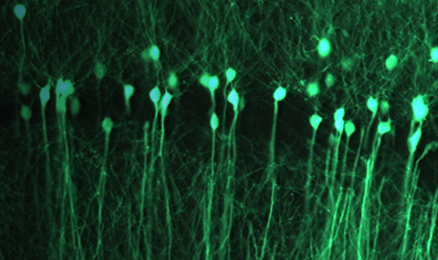
The dynamic internal representation of past experience is a defining feature of ‘Memory’. This internal representation of information is thought to be carried out by the activity of specific ensembles of neurons in specialized brain circuits, such as the hippocampus. Following initial learning, memory-associated ensemble activity undergoes a time- and experience-dependent evolution. Biological processes at various levels of organization (molecular, cellular, circuit) determine the fate of stored memories: some memories are lost very rapidly, while others can persist for years. Research in our lab focuses on the mechanisms that underlie memory’s dynamic persistence: How do memory representations evolve as a function of learning? How do memory representations change over periods of weeks and months? How do age-related neurological disorders degrade memory representations? How do different plasticity mechanisms contribute to processing of memory information?
To address these questions we investigate how memory information is encapsulated in the codes that are generated by neuronal ensembles in brain circuits that are important for memory processing. We follow memory representations over long periods of time (days, weeks, and months) using novel optical imaging methods. These methods allows for longitudinal optical recording of neuronal activity from large neuronal populations (of > 1,000 neurons) in memory circuits of freely behaving rodents. We combine imaging methodologies with genetic tools for manipulating specific molecular pathways or spiking activity in defined cell types, behavioral assays for spatial learning and long-term memory, and computational methods for analyzing data from large-scale neuronal populations.
Our current research centers on neural coding in the hippocampus, a brain structure that is crucial for spatial navigation and for the formation and processing of memories for places and events. We are studying the roles of plasticity mechanisms, such as adult neurogenesis in the dentate gyrus, in processing of long-term spatial memory, and how hippocampal representation of space is linked to long-term spatial memory under both normal and pathological conditions.
|
Chronic Ca2+-imaging in the hippocampus of a freely behaving mouse – a new approach to study neural coding of long-term memory. A video showing a mouse exploring a circular arena (left panel) and the simultaneously acquired brain-imaging data of CA1 pyramidal cell Ca2+ activity, displayed as relative changes in fluorescence (deltaF/F) (right panel). 705 pyramidal cells were identified in the total data set. The Ca2+-imaging frame rate was 20 Hz, but these data are shown down-sampled to 5 Hz to aid visualization of the Ca2+ transients. The video playback rate is sped up four-fold from how the events actually occurred. Taken from: Nature Neuroscience 2013. |


