Skip to main content
Skip to navigation
Skip to main content
Open accessibility toolbar
Accessibility
Grayscale
Highlight Links
Contrast
Negative Contrast
Increase font
Decrease font
Stop animations
Reset All
Disclaimer
Accessibility arrangements
Immunology & Regenerative Biology
Benny Geiger's Lab
Cellular Communication
Menu
Main navigation
Home
Research
Publications
Group
Gallery
Collaborations
Contact
Breadcrumb
Home
Gallery
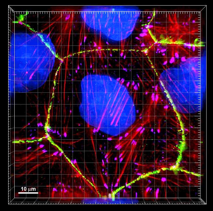
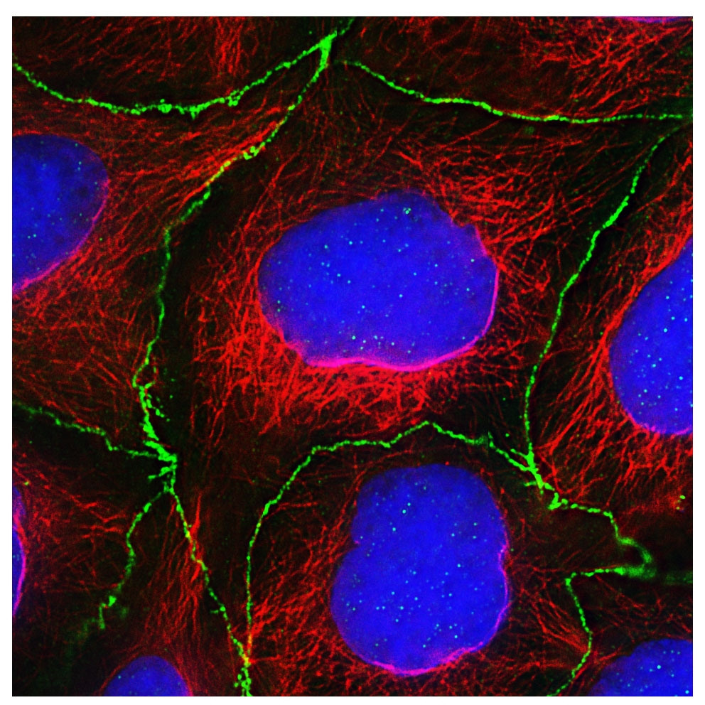
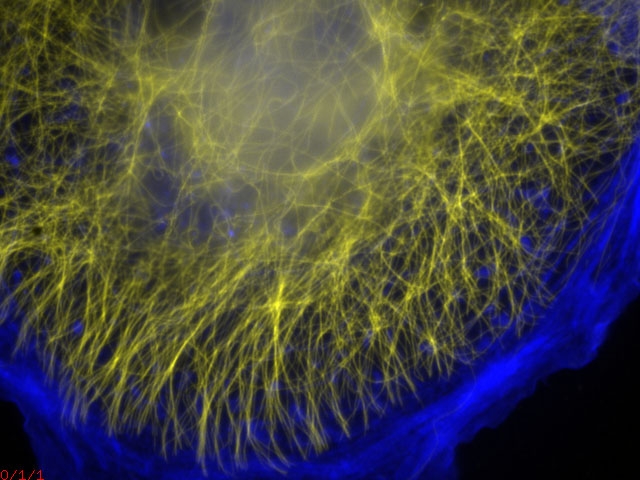
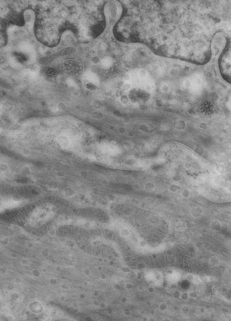
Lab images
Lab images