Research

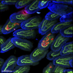
macrophages (red) in small intestinal villi, vasculature (green)

Macrophages - intestine / brain

Tissue macrophages are strategically positioned throughout the organism. As professional phagocytes, they ingest and degrade debris and foreign material, including pathogens, and orchestrate inflammatory processes. Macrophages can be generated from three distinct sources: two early, currently considered transient, hematogenic waves initiated in the yolk sac that seed tissues prenatally, and a pathway involving hematopoietic stem cells and mature blood monocytes, that persists throughout adult life. Most macrophage compartments are established prenatally, and develop independently from each other in their respective host tissue. These cells locally acquire distinct epigenomic and transcriptomic identities, through the imprint influence of the respective microenvironment. While recent studies have revealed critical contributions of tissue macrophages to organ development and homeostasis, specific involvement of discrete subpopulations in the maintenance of the healthy balanced steady-state remain however to be defined. Using the mouse as a model, we aim to define functional contributions of specific tissue macrophages to physiology and pathophysiology.

Monocytes

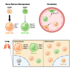
Monocytes are mononuclear phagocytes that circulate in the blood. One monocyte subset seems dedicated to patrol the inner vessel wall and surveille it for integrity. A second population, defined as classical monocytes and CD14+ cells and Ly6C+ cells in man and mouse respectively, is highly dynamic and short-lived. These cells can be recruited to sites of inflammation and injury where they can give rise to monocyte-derived DC and macrophages. Emerging data suggest that classical monocytes themselves comprise neutrophil-like and DC-like populations. However, differential functional contributions of these cells to physiology and pathophysiology, as well as their relation to tissue-resident moDC and macrophages remain to be defined. We approach this question in the mouse using adoptive transfers and fate mapping strategies. 

Dendritic cells

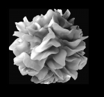
Classical Dendritic cells (cDC) are migratory myeloid cells with an unrivaled potential to stimulate naïve T cells. Adoptive transfer experiments have established that cDC are derived from dedicated DC precursors which differentiate in tissue context into distinct DC subsets, including XCR1+ (CD8a+) cDC and CD11b+ DC. Functional differences between these subsets remain incompletely understood, as is their relation to monocyte-derived DC that complement the cDC compartment during inflammation.