
It’s common high school knowledge that almost all organisms inherit their mitochondria from the maternal parent. But what is the fate of the paternal mitochondria? And why are they prevented from getting passed on to their offspring after fertilization? The answers for these questions have been shrouded in mystery for several decades. Whereas some reports supported a passive dilution theory of the paternal mitochondria in excess copy number of the maternal mitochondria, others implied the involvement of active destruction mechanisms.
We investigate the fate of the paternal mitochondria after fertilization in Drosophila. Interestingly, we found that the paternal mitochondrial derivative is targeted for rapid destruction by mechanisms originated in the egg. In particular, we identified egg-derived unique vacuoles of dual pathway (a common endocytic and autophagic pathway), which associate with the sperm plasma membrane upon fertilization and promote the destruction of the mitochondria. This system allows us to investigate the mechanisms and principles of organelle turnover under physiological conditions, as well as study the basis for the selectivity of this process (i.e. how the egg “differentiates” between its own mitochondria and the paternal mitochondria).
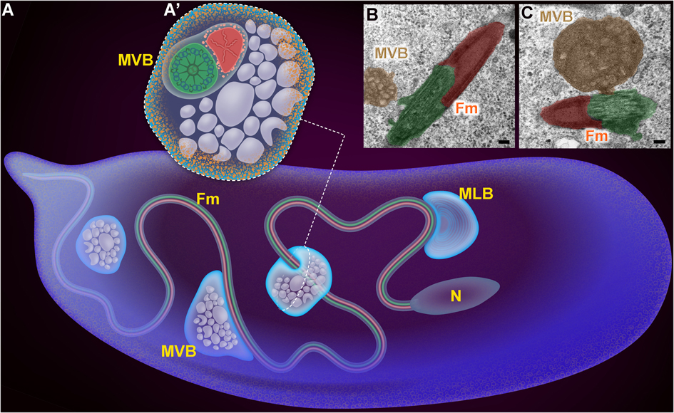
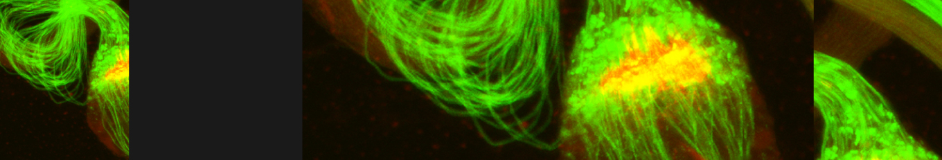
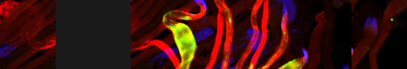
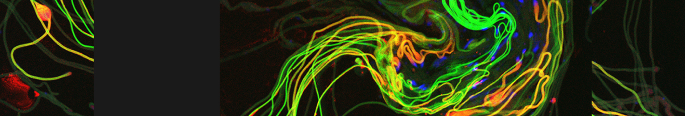
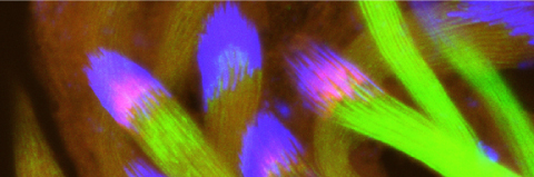
Egg-derived active mechanisms eliminate the paternal mitochondria after fertilization in Drosophila (A) Schematic representation of an early fertilized Drosophila egg. The penetrating sperm is mainly composed of a needle-shape nucleus (N) and an almost 2 mm long flagellum (Fm), consisting of two major elongated organelles, an axoneme (green) and a mitochondrial derivative (red). Soon after fertilization, a network of multivesicular body (MVB)-like vesicles originating in the egg and displaying molecular features of both the endocytic and autophagic pathways, approach and associate with the sperm flagellum, and are involved in the initial recognition and disintegration of the sperm plasma membrane after fertilization. MLB, multilamellar body. (A’) An illustration of a cross-section through one of these MVBs, depicting ubiquitinated proteins (pink dots) on the surface of the mitochondrial derivative (red) and the localization of the LC3/Atg8-based fluorescent autophagic reporter at the circumference of these vesicles (light orange dots). (B, C) Electron micrographs of cross-sections through early fertilized eggs. MVBs approach (B) and associate (C) with the plasma membrane surrounding the flagellum (Fm). The colors indicated (MVBs, bronze; mitochondrial derivative, red; axoneme, green) were manually added using Adobe Photoshop to correspond to the colored schematic organelles in (A). Scale bars represent 100 nm.