Research

What determines cell size? Studies of the genetic disease ARO

We have constructed mice carrying the R51Q mutation in sorting nexin 10 (SNX10), which was suggested to cause the lethal genetic disease autosomal recessive osteopetrosis (ARO) in humans. Mice homozygous for this mutation are indeed osteopetrotic, but their osteoclasts exhibit a highly unsusual phenotype: the cells fuse constantly and repeatedly and form osteoclasts that are 1-2 orders of magnitude larger than wild-type cells. This result indicates that the upper size limit of osteoclasts is determined by an active, genetically-regulated mechanism in which SNX10 plays a significant role, and enables addressing basic questions in the biology of cell fusion.

What drives osteoclasts to begin to fuse?

Monocytes and macrophages differentiate into bone-resorbing osteoclasts (OCLs) under the direction of the cytokines M-CSF and RANKL. It is believed that RANKL induces a small fraction of monocytes to become “Founder” cells, which have the capacity to initiate cell-cell fusion. The remaining cells are “Follower” cells, which can fuse with Founder cells but cannot initiate the process themselves. Both classes of cells are morphologically identical, and the molecular differences between them are unknown. We are devising methods to isolate Founder cells and to study the molecular basis for their ability to initiate fusion in order to better understand the initial stages of OCL formation.

Tyrosine Phosphatases in Bone Biology

Osteoclasts are hematopoietically-derived cells whose function is to degrade bone. The production and then function of these cells involves complex cell-signaling processes, in which protein tyrosine kinases and phosphatases play central roles. We are using knockout and knock-in mice of our production, as well as primary osteoclasts derived from them, to provide detailed molecular mechanisms of how several tyrosine phosphatases, such as PTPRE, PTPRO, adn PTPRJ, function in osteoclasts.


Protein Tyrosine Phosphatases

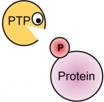
Phosphorylation of tyrosine residues in proteins is a major mechanism for regulating their structure and function. Phosphorylation is reversible and is regulated by the generically opposing activities of tyrosine kinases and tyrosine phosphatases (PTPs).

Over 100 PTP genes are known. The products of these genes exhibit substrate specificity that ranges from exclusively phospho-tyrosine through phospho-serine and –threonine and up to lipids and RNA. These enzymes are classified as PTPs since their catalytic mechanism is similar and is characteristic of the tyrosine-specific, so-called “classical” PTP family, which was the first to be discovered by Nicholas Tonks and Edmond Fischer in 1988.

A very large number of studies has established that dysregulation of protein phosphorylation can lead to serious disease, including cancer, metabolic syndrome, neural dysfunction, and others. Molecules that regulate these processes are therefore critical modulators of cellular function, as has been shown by increasing development and use of modulators of tyrosine kinase activity as drugs for treating disease. Tyrosine phosphatases play roles of similar importance in these processes, and their specificity in vivo is high.  Since they are entirely distinct from tyrosine kinases, study of PTPs and their molecular roles in physiological processes can shed new light on the molecular details of these processes and suggest novel ways to intervene in them for therapeutic gain.