Publications
2024
-
(2024) Nature Chemical Biology. Abstract
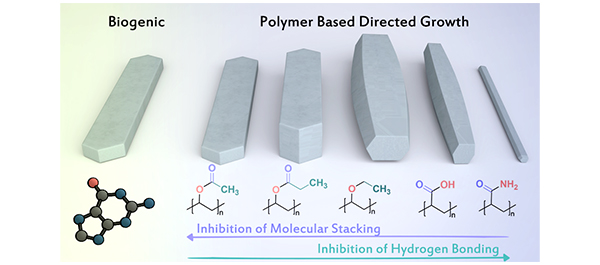
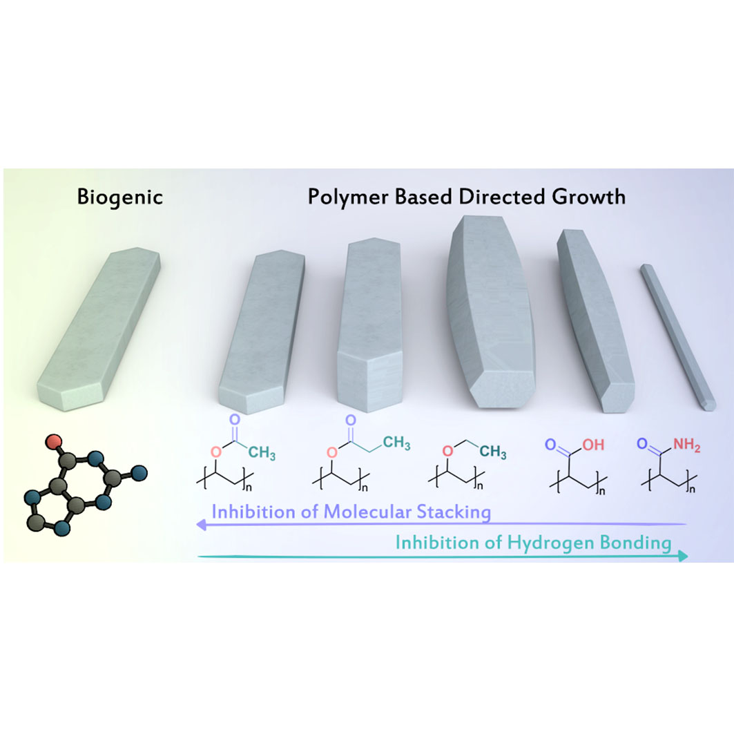
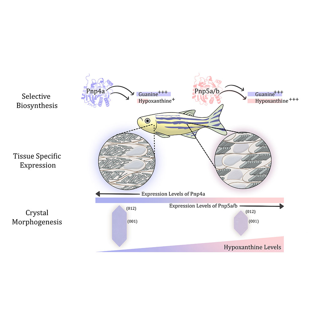
Organisms evolve mechanisms that regulate the properties of biogenic crystals to support a wide range of functions, from vision and camouflage to communication and thermal regulation. Yet, the mechanism underlying the formation of diverse intracellular crystals remains enigmatic. Here we unravel the biochemical control over crystal morphogenesis in zebrafish iridophores. We show that the chemical composition of the crystals determines their shape, particularly through the ratio between the nucleobases guanine and hypoxanthine. We reveal that these variations in composition are genetically controlled through tissue-specific expression of specialized paralogs, which exhibit remarkable substrate selectivity. This orchestrated combination grants the organism with the capacity to generate a broad spectrum of crystal morphologies. Overall, our findings suggest a mechanism for the morphological and functional diversity of biogenic crystals and may, thus, inspire the development of genetically designed biomaterials and medical therapeutics. (Figure presented.).
-
(2024) BioRxiv. 2024.07.20. Abstract
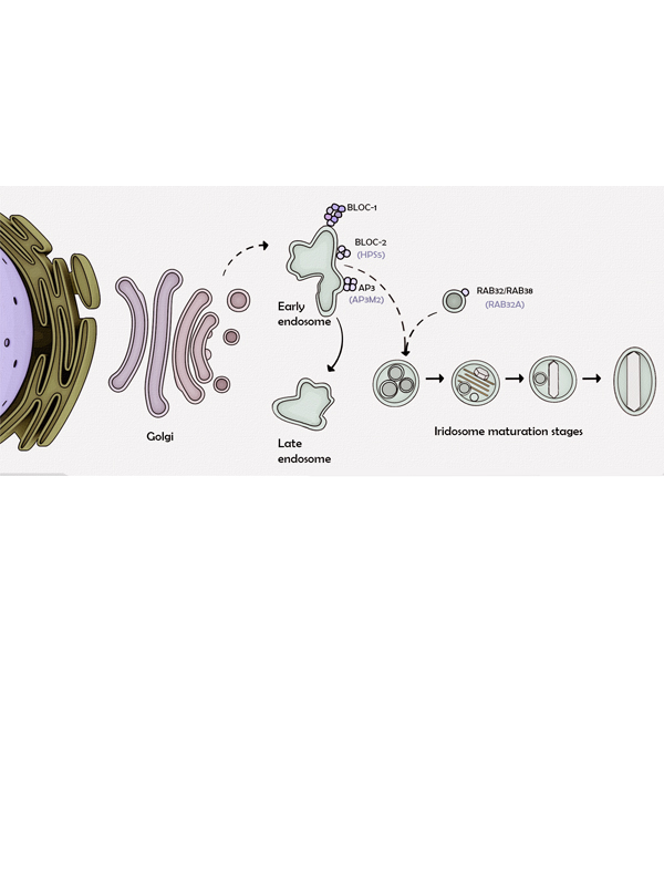
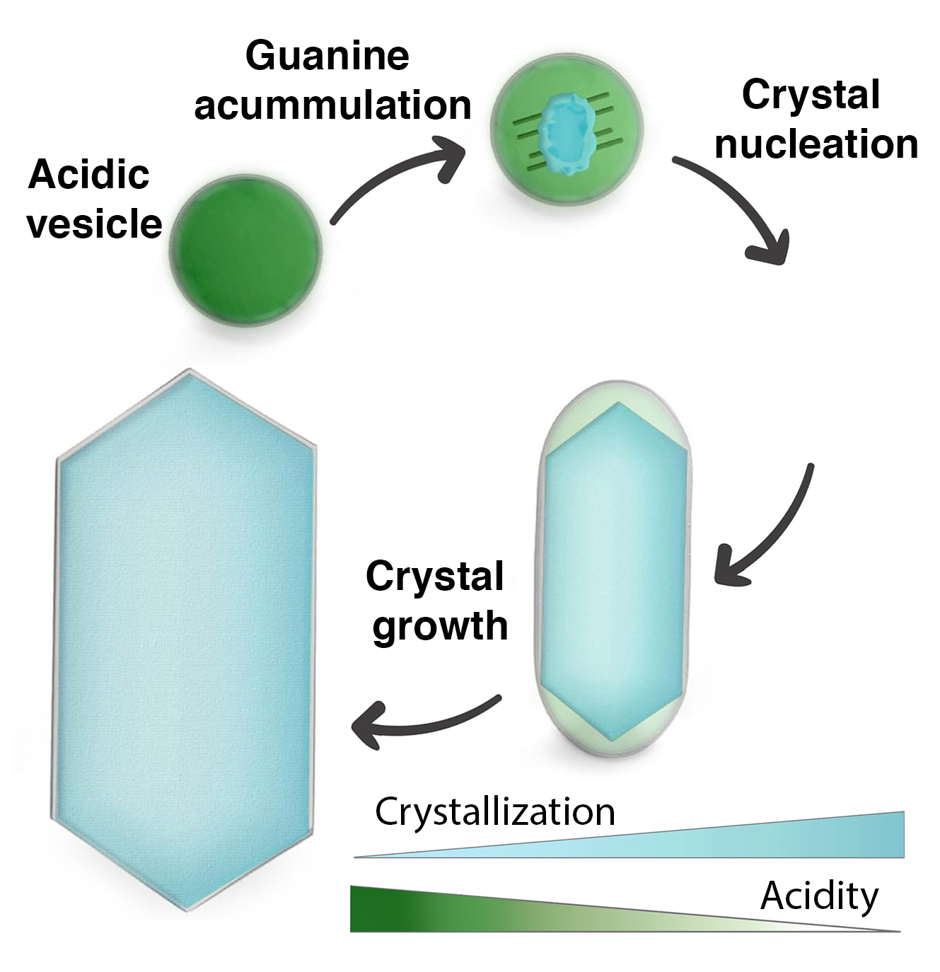
Many animals exhibit remarkable colors produced by the constructive interference of light reflected from arrays of intracellular guanine crystals. These systems are utilized for various purposes, including vision, camouflage, communication, and thermal regulation. Each guanine crystal forms within a membrane-bound organelle called an iridosome, where precise control over crystal formation occurs. While the presence of guanine crystals in iridosomes is well-documented, the mechanisms facilitating the accumulation of water-insoluble guanine and driving its crystallization remain unclear. Here, we employ advanced imaging and spectroscopy techniques to characterize the maturation of iridosomes in zebrafish iridophores during development. Using cryo-electron microscopy, we found that amorphous guanine accumulates in early-stage iridosomes. Synchrotron-based soft X-ray microscopy studies revealed that, unlike mature crystals, the accumulated guanine is initially in its protonated state. Live imaging with a pH sensor demonstrated that early-stage iridosomes are acidic and that their pH gradually approaches neutrality during maturation. Additionally, the application of a V-ATPase inhibitor reduced the acidity of iridosomes and significantly decreased crystal formation, suggesting the involvement of V-ATPase in regulating the organelle pH. Our findings reveal new insights into the molecular mechanisms facilitating guanine accumulation and crystallization within iridosomes, emphasizing the pivotal role of pH alternations in the precise formation of biogenic crystals.Competing Interest StatementThe authors have declared no competing interest.
-
(2024) Proceedings of the National Academy of Sciences. 121, 23, e230853112. Abstract
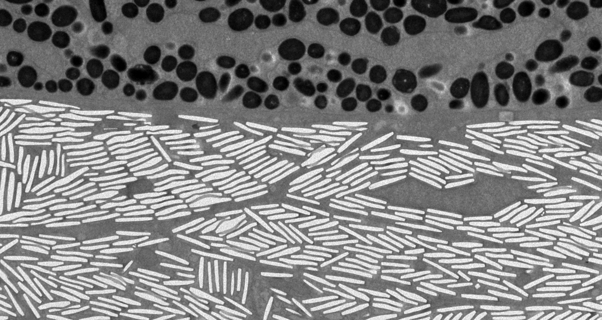
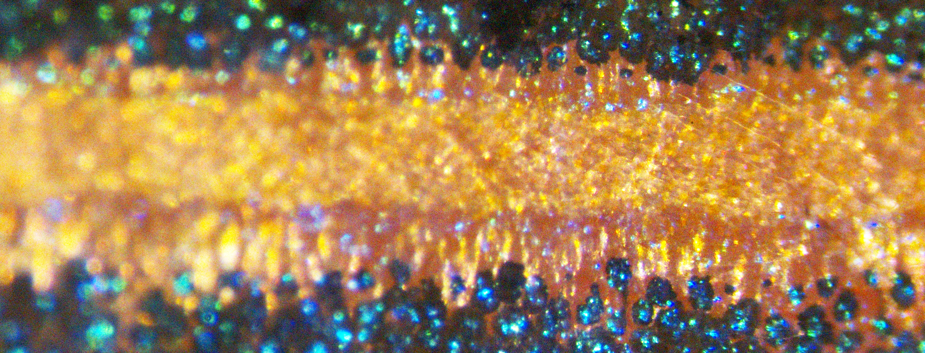
Many animals exhibit remarkable colors that are produced by the constructive interference of light reflected from arrays of intracellular guanine crystals. These animals can fine-tune their crystal-based structural colors to communicate with each other, regulate body temperature, and create camouflage. While it is known that these changes in color are caused by changes in the angle of the crystal arrays relative to incident light, the cellular machinery that drives color change is not understood. Here, using a combination of 3D focused ion beam scanning electron microscopy (FIB-SEM), micro-focused X-ray diffraction, superresolution fluorescence light microscopy, and pharmacological perturbations, we characterized the dynamics and 3D cellular reorganization of crystal arrays within zebrafish iridophores during norepinephrine (NE)-induced color change. We found that color change results from a coordinated 20° tilting of the intracellular crystals, which alters both crystal packing and the angle at which impinging light hits the crystals. Importantly, addition of the dynein inhibitor dynapyrazole-a completely blocked this NE-induced red shift by hindering crystal dynamics upon NE addition. FIB-SEM and microtubule organizing center (MTOC) mapping showed that microtubules arise from two MTOCs located near the poles of the iridophore and run parallel to, and in between, individual crystals. This suggests that dynein drives crystal angle change in response to NE by binding to the limiting membrane surrounding individual crystals and walking toward microtubule minus ends. Finally, we found that intracellular cAMP regulates the color change process. Together, our results provide mechanistic insight into the cellular machinery that drives structural color change.
2022
-
(2022) Journal of the American Chemical Society. 144, 49, p. 22440-22445 Abstract
Controlling the morphology of crystalline materials is challenging, as crystals have a strong tendency toward thermodynamically stable structures. Yet, organisms form crystals with distinct morphologies, such as the plate-like guanine crystals produced by many terrestrial and aquatic species for light manipulation. Regulation of crystal morphogenesis was hypothesized to entail physical growth restriction by the surrounding membrane, combined with fine-tuned interactions between organic molecules and the growing crystal. Using cryo-electron tomography of developing zebrafish larvae, we found that guanine crystals form via templated nucleation of thin leaflets on preassembled scaffolds made of 20-nm-thick amyloid fibers. These leaflets then merge and coalesce into a single plate-like crystal. Our findings shed light on the biological regulation of crystal morphogenesis, which determines their optical properties.
2020
-
(2020) Nature Communications. 11, 1, 6391. Abstract
Skin color patterns are ubiquitous in nature, impact social behavior, predator avoidance, and protection from ultraviolet irradiation. A leading model system for vertebrate skin patterning is the zebrafish; its alternating blue stripes and yellow interstripes depend on light-reflecting cells called iridophores. It was suggested that the zebrafishs color pattern arises from a single type of iridophore migrating differentially to stripes and interstripes. However, here we find that iridophores do not migrate between stripes and interstripes but instead differentiate and proliferate in-place, based on their micro-environment. RNA-sequencing analysis further reveals that stripe and interstripe iridophores have different transcriptomic states, while cryogenic-scanning-electron-microscopy and micro-X-ray diffraction identify different crystal-arrays architectures, indicating that stripe and interstripe iridophores are different cell types. Based on these results, we present an alternative model of skin patterning in zebrafish in which distinct iridophore crystallotypes containing specialized, physiologically responsive, organelles arise in stripe and interstripe by in-situ differentiation.
2019
-
(2019) Development. 146, 23, dev177790. Abstract
To maintain body homeostasis, endocrine systems must detect and integrate blood-borne peripheral signals. This is mediated by fenestrae, specialized permeable pores in the endothelial membrane. Plasmalemma vesicle-associated protein (Plvap) is located in the fenestral diaphragm and is thought to play a role in the passage of proteins through the fenestrae. However, this suggested function has yet to be demonstrated directly. We studied the development of fenestrated capillaries in the hypophysis, a major neuroendocrine interface between the blood and brain. Using a transgenic biosensor to visualize the vascular excretion of the genetically tagged plasma protein DBP-EGFP, we show that the developmental acquisition of vascular permeability coincides with differential expression of zebrafish plvap orthologs in the hypophysis versus brain. Ultrastructural analysis revealed that plvapb mutants display deficiencies in fenestral diaphragms and increased density of hypophyseal fenestrae. Measurements of DBP-EGFP extravasation in plvapb mutants provided direct proof that Plvap limits the rate of blood-borne protein passage through fenestrated endothelia. We present the regulatory role of Plvap in the development of blood-borne protein detection machinery at a neuroendocrine interface through which hormones are released to the general circulation.
-
(2019) Proceedings of the National Academy of Sciences of the United States of America. 116, 24, p. 11806-11811 Abstract
Understanding genetic and cellular bases of adult form remains a fundamental goal at the intersection of developmental and evolutionary biology. The skin pigment cells of vertebrates, derived from embryonic neural crest, are a useful system for elucidating mechanisms of fate specification, pattern formation, and how particular phenotypes impact organismal behavior and ecology. In a survey of Danio fishes, including the zebrafish Danio rerio, we identified two populations of white pigment cellsleucophoresone of which arises by transdifferentiation of adult melanophores and another of which develops from a yelloworange xanthophore or xanthophore-like progenitor. Single-cell transcriptomic, mutational, chemical, and ultrastructural analyses of zebrafish leucophores revealed cell-typespecific chemical compositions, organelle configurations, and genetic requirements. At the organismal level, we identified distinct physiological responses of leucophores during environmental background matching, and we showed that leucophore complement influences behavior. Together, our studies reveal independently arisen pigment cell types and mechanisms of fate acquisition in zebrafish and illustrate how concerted analyses across hierarchical levels can provide insights into phenotypes and their evolution.
2018
-
(2018) Advanced Materials. 30, 41, 1800006. Abstract
Vision mechanisms in animals, especially those living in water, are diverse. Many eyes have reflective elements that consist of multilayers of nanometer-sized crystalline plates, composed of organic molecules. The crystal multilayer assemblies owe their enhanced reflectivity to the high refractive indices of the crystals in preferred crystallographic directions. The high refractive indices are due to the molecular arrangements in their crystal structures. Herein, data regarding these difficult-to-characterize crystals are reviewed. This is followed by a discussion on the function of these crystalline assemblies, especially in visual systems whose anatomy has been well characterized under close to in vivo conditions. Three test cases are presented, and then the relations between the reflecting crystalline components and their functions, including the relations between molecular structure, crystal structure, and reflecting properties are discussed. Some of the underlying mechanisms are also discussed, and finally open questions in the field are identified.
-
(2018) ACS Central Science. 4, 8, p. 1031-1036 Abstract
Organic crystals are of primary importance in pharmaceuticals, functional materials, and biological systems; however, organic crystallization mechanisms are not well-understood. It has been recognized that "nonclassical" organic crystallization from solution involving transient amorphous precursors is ubiquitous. Understanding how these precursors evolve into crystals is a key challenge. Here, we uncover the crystallization mechanisms of two simple aromatic compounds (perylene diimides), employing direct structural imaging by cryogenic electron microscopy. We reveal the continuous evolution of density, morphology, and order during the crystallization of very different amorphous precursors (well-defined aggregates and diffuse dense liquid phase). Crystallization starts from initial densification of the precursors. Subsequent evolution of crystalline order is gradual, involving further densification concurrent with optimization of molecular ordering and morphology. These findings may have implications for the rational design of organic crystals.
-
(2018) Advanced Science. 5, 8, 1800338. Abstract
Many marine organisms have evolved a reflective iris to prevent unfocused light from reaching the retina. The fish iris has a dual function, both to camouflage the eye and serving as a light barrier. Yet, the physical mechanism that enables this dual functionality and the benefits of using a reflective iris have remained unclear. Using synchrotron microfocused diffraction, cryo-scanning electron microscopy imaging, and optical analyses on zebrafish at different stages of development, it is shown that the complex optical response of the iris is facilitated by the development of high-order organization of multilayered guanine-based crystal reflectors and pigments. It is further demonstrated how the efficient light reflector is established during development to allow the optical functionality of the eye, already at early developmental stages.
-
(2018) Journal of the Royal Society Interface. 15, 139, 20170930. Abstract
This study investigates the structural basis for the red, silver and black coloration of the theridiid spider, Phoroncidia rubroargentea (Berland, 1913) from Madagascar. Specimens of this species can retain their colour after storage in ethanol for decades, whereas most other brightly pigmented spider specimens fade under identical preservation conditions. Using correlative optical, structural and chemical analysis, we identify the colour-generating structural elements and characterize their optical properties. The prominent silvery appearance of the spider's abdomen results from regularly arranged guanine microplatelets, similar to those found in other spiders and fish. The microplatelets are composed of a doublet structure twinned about the [02(1) over bar] axis, as suggested by electron diffraction. The red coloration originates from chambered microspheres (approx. 1 mu m in diameter), which contain structured fluorescent material. Co-localization of the red microparticles on top of the reflective guanine microplatelets appears to enhance the red coloration. The spider's thick cuticular layer, which encases its abdomen, varies in its optical properties, being transparent in regions where only guanine reflectors are present, and tanned, exhibiting light absorption where the red microspheres are found. Moreover, colour degradation in some preserved spider specimens that had suffered damage to the cuticular layer suggests that this region of the exoskeleton may play an important role in the stabilization of the red coloration.
2017
-
(2017) Science. 358, 6367, p. 1172-1175 Abstract
Scallops possess a visual system comprising up to 200 eyes, each containing a concave mirror rather than a lens to focus light. The hierarchical organization of the multilayered mirror is controlled for image formation, from the component guanine crystals at the nanoscale to the complex three-dimensional morphology at the millimeter level. The layered structure of the mirror is tuned to reflect the wavelengths of light penetrating the scallop's habitat and is tiled with a mosaic of square guanine crystals, which reduces optical aberrations. The mirror forms images on a double-layered retina used for separately imaging the peripheral and central fields of view. The tiled, off-axis mirror of the scallop eye bears a striking resemblance to the segmented mirrors of reflecting telescopes.
-
(2017) ANGEWANDTE CHEMIE-INTERNATIONAL EDITION. 56, 32, p. 9420-9424 Abstract
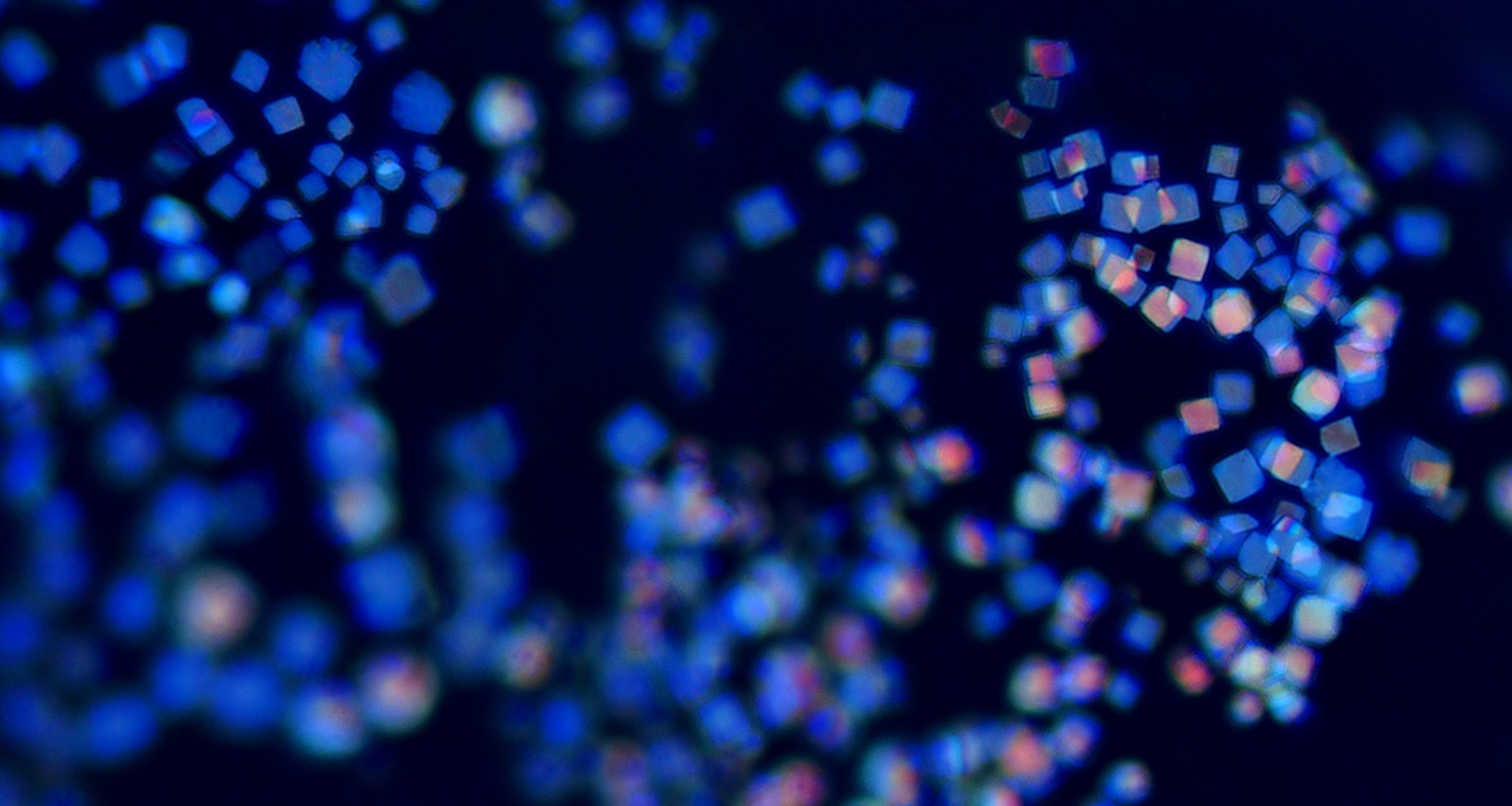
Guanine crystals are widely used in nature as components of multilayer reflectors. Guanine-based reflective systems found in the copepod cuticle and in the mirror of the scallop eye are unique in that the multilayered reflectors are tiled to form a contiguous packed array. In the copepod cuticle, hexagonal crystals are closely packed to produce brilliant colors. In the scallop eye, square crystals are tiled to obtain an image-forming reflecting mirror. The tiles are about 1 mm in size and 70 nm thick. According to analysis of their electron diffraction patterns, the hexagon and square tiles are not single crystals. Rather, each tile type is a composite of what appears to be three crystalline domains differently oriented and stacked onto one another, achieved through a twice-repeated twinning about their and crystal axes, respectively. By these means, the monoclinic guanine crystal mimics higher symmetry hexagonal and tetragonal structures to achieve unique morphologies.
-
(2017) Advanced Functional Materials. 27, 6, 1603514. Abstract
Guanine crystals are widely used in nature to manipulate light. The first part of this feature article explores how organisms are able to construct an extraordinary array of optical "devices" including diffuse scatterers, broad-band and narrowband reflectors, tunable photonic crystals, and image-forming mirrors by varying the size, morphology, and arrangement of guanine crystals. The second part presents an overview of some of the properties of crystalline guanine to explain why this material is ideally suited for such optical applications. The high reflectivity of many natural optical systems ultimately derives from the fact that guanine crystals have an extremely high refractive index-a product of its anisotropic crystal structure comprised of densely stacked H-bonded layers. In order to optimize their reflectivity, many organisms exert exquisite control over the crystal morphology, forming plate-like single crystals in which the high refractive index face is preferentially expressed. Guanine-based optics are used in a wide range of biological functions such as in camouflage, display, and vision, and exhibit a degree of versatility, tunability, and complexity that is difficult to incorporate into artificial devices using conventional engineering approaches. These biological systems could inspire the next generation of advanced optical materials.
-
(2017) Advanced Optical Materials. 5, 1, 1600617. Abstract
The bacteria-based photonic device is analogous to liquid-crystal-based devices where the liquid crystals and an electric field are replaced by magnetic bacteria and a magnetic field, respectively. The controlled orientation of the bacteria in the device is used to tune the intensity and the polarization of the light.
2016
-
(2016) Crystal Growth & Design. 16, 9, p. 4975-4980 Abstract
Anhydrous guanine crystals are among the most widespread organic crystals used by organisms to produce structural colors. The main advantage of guanine is its exceptionally high refractive index in the reflecting direction (∼1.8). For the same reason, guanine is a promising candidate material for a variety of different optical applications. Crystallization of guanine is challenging and usually involves using polar aprotic organic solvents such as dimethyl sulfoxide (DMSO). Here, we show that the crystallization of guanine from aqueous solutions is possible under conditions that provide control over crystal polymorphism and size. Using this approach we were able produce large crystals of the elusive guanine monohydrate phase. We were also able to rationalize the formation of the different phases obtained as a function of which tautomer of guanine is stable in solutions of varying pH.
-
(2016) Nature Communications. 7, 11592. Abstract
The author Hagai Cohen is incorrectly omitted from the list of corresponding authors. The corresponding authors are Peter Rez and Hagai Cohen. The correct information for correspondence is: Correspondence and requests for materials should be addressed to P.R. (Peter.Rez@asu.edu) or to H.C. (Hagai.Cohen@Weizmann.ac.il).
-
(2016) Advanced Functional Materials. 26, 9, p. 1393-1399 Abstract
Light-induced tunable photonic systems are rare in nature, and generally beyond the state-of-the-art in artificial systems. Sapphirinid male copepods produce some of the most spectacular colors in nature. The male coloration, used for communication purposes, is structural and is produced from ordered layers of guanine crystals separated by cytoplasm. It is generally accepted that the colors of the males are related to their location in the epipelagic zone. By combining correlative reflectance and cryoelectron microscopy image analyses, together with optical time lapse recording and transfer matrix modeling, it is shown that male sapphirinids have the remarkable ability to change their reflectance spectrum in response to changes in the light conditions. It is also shown that this color change is achieved by a change in the thickness of the cytoplasm layers that separate the guanine crystals. This change is reversible, and is both intensity and wavelength dependent. This capability provides the male with the ability to efficiently reflect light under certain conditions, while remaining transparent and hence camouflaged under other conditions. These copepods can thus provide inspiration for producing synthetic tunable photonic arrays.
-
(2016) Nature Communications. 7, 10945. Abstract
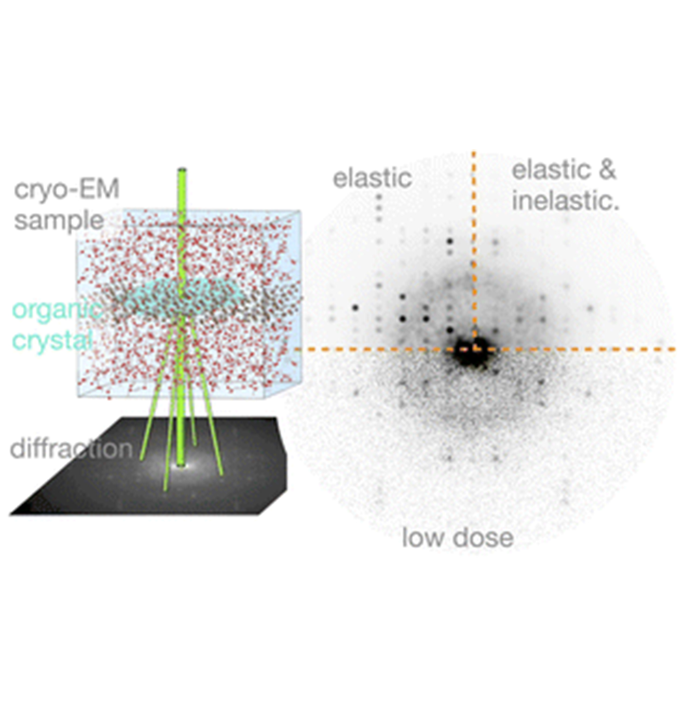
Vibrational spectroscopy in the electron microscope would be transformative in the study of biological samples, provided that radiation damage could be prevented. However, electron beams typically create high-energy excitations that severely accelerate sample degradation. Here this major difficulty is overcome using an 'aloof' electron beam, positioned tens of nanometres away from the sample: high-energy excitations are suppressed, while vibrational modes of energies
2015
-
(2015) Chemistry of Materials. 27, 24, p. 8289-8297 Abstract
Living organisms display a spectrum of wondrous colors, which can be produced by pigmentation, structural coloration, or a combination of the two. A relatively well-studied system, which produces colors via an array of alternating anhydrous guanine crystals and cytoplasm, is responsible for the metallic luster of many fish. The structure of biogenic anhydrous guanine was so far believed to be the same as that of the synthetic one, a monoclinic polymorph (denoted as α). Here we re-examine the structure of biogenic guanine, using detailed experimental X-ray and electron diffraction data, exposing troublesome inconsistencies, namely, a "guanigma". To address this, we sought alternative candidate polymorphs using symmetry and packing considerations and then utilized first-principles calculations to determine whether the selected candidates could be energetically stable. We identified theoretically a different monoclinic polymorph (denoted as β), were able to synthesize it, and confirmed using X-ray diffraction that it is this polymorph that occurs in biogenic samples. However, the electron diffraction data were still not consistent with this polymorph but rather with a theoretically generated orthorhombic polymorph (denoted as γ). This apparent inconsistency was resolved by showing how the electron diffraction pattern could be affected by crystal structural faults composed of offset molecular layers.
-
(2015) Journal of the American Chemical Society. 137, 26, p. 8408-8411 Abstract
Males of sapphirinid copepods use regularly alternating layers of hexagonal-shaped guanine crystals and cytoplasm to produce spectacular structural colors. In order to understand the mechanism by which the different colors are produced, we measured the reflectance of live individuals and then characterized the organization of the crystals and the cytoplasm layers in the same individuals using cryo-SEM. On the basis of these measurements, we calculated the expected reflectance spectra and found that they are strikingly similar to the measured ones. We show that variations in the cytoplasm layer thickness are mainly responsible for the different reflected colors and also that the copepod color strongly depends on the angular orientation relative to the incident light, which can account for its appearance and disappearance during spiral swimming in the natural habitat.
-
The Mechanism of Color Change in the Neon Tetra Fish: A Light-Induced Tunable Photonic Crystal Array(2015) Angewandte Chemie (International ed. in English). 54, 42, p. 12426-12430 Abstract
The fresh water fish neon tetra has the ability to change the structural color of its lateral stripe in response to a change in the light conditions, from blue-green in the light-adapted state to indigo in the dark-adapted state. The colors are produced by constructive interference of light reflected from stacks of intracellular guanine crystals, forming tunable photonic crystal arrays. We have used micro X-ray diffraction to track in time distinct diffraction spots corresponding to individual crystal arrays within a single cell during the color change. We demonstrate that reversible variations in crystal tilt within individual arrays are responsible for the light-induced color variations. These results settle a long-standing debate between the two proposed models, the "Venetian blinds" model and the "accordion" model. The insight gained from this biogenic light-induced photonic tunable system may provide inspiration for the design of artificial optical tunable systems.
-
(2015) Acta crystallographica. Section E, Crystallographic communications. 71, 3, p. 281-283 Abstract
In the title compound, disodium 2-amino-6-oxo-6,7-dihydro-1H-purine-1,7-diide heptahydrate, 2Na+·C5H3N5O2-·7H2O, the structure is composed of alternating (100) layers of guanine molecules and hydrated Na+ ions. Within the guanine layer, the molecules are arranged in centrosymmetric pairs, with a partial overlap between the guanine rings. In this compound, guanine exists as the amino-keto tautomer from which deprotonation from N1 and N7 has occurred (purine numbering). There are no direct interactions between the Na+ cations and the guanine anions. Guanine molecules are linked to neighboring water molecules by O - H⋯N and O - H⋯O hydrogen bonds into a network structure.
2014
-
(2014) Journal of the American Chemical Society. 136, 49, p. 17236-17242 Abstract
Fish have evolved biogenic multilayer reflectors composed of stacks of intracellular anhydrous guanine crystals separated by cytoplasm, to produce the silvery luster of their skin and scales. Here we compare two different variants of the Japanese Koi fish; one of them with enhanced reflectivity. Our aim is to determine how biology modulates reflectivity, and from this to obtain a mechanistic understanding of the structure and properties governing the intensity of silver reflectance. We measured the reflectance of individual scales with a custom-made microscope, and then for each individual scale we characterized the structure of the guanine crystal/cytoplasm layers using high-resolution cryo-SEM. The measured reflectance and the structural-geometrical parameters were used to calculate the reflectance of each scale, and the results were compared to the experimental measurements. We show that enhanced reflectivity is obtained with the same basic guanine crystal/cytoplasm stacks, but the structural arrangement between the stack, inside the stacks, and relative to the scale surface is varied when reflectivity is enhanced. Finally, we propose a model that incorporates the basic building block parameters, the crystal orientation inside the tissue, and the resulting reflectance and explains the mechanistic basis for reflectance enhancement.
2013
-
(2013) Angewandte Chemie (International ed.). 52, 18, p. 4867-4870 Abstract
An ugly duckling grows into a swan: Many organisms grow their crystalline mineral phases through the secondary nucleation of nanospheres made of an amorphous precursor phase. Stable amorphous calcium carbonate biominerals were used to induce a similar transformation in vitro. The amorphous nanospheres underwent a solidphase transformation that resulted in highly ordered calcite crystals composed of aggregated particles.
-
(2013) ANGEWANDTE CHEMIE-INTERNATIONAL EDITION. 52, 1, p. 388-391 Abstract
Starting from disorder: Anhydrous guanine crystals compose the photonic arrays responsible for the skin and scale iridescence found in Japanese Koi fish. These guanine crystals were found to form in intracellular vesicles (see picture) through an amorphous precursor phase. A combined cryo-SEM and synchrotron radiation X-ray diffraction study showed the evolution of the crystals in great detail.
2011
-
(2011) Journal of Structural Biology. 174, 3, p. 527-535 Abstract
Bone is the most widespread mineralized tissue in vertebrates and its formation is orchestrated by specialized cells - the osteoblasts. Crystalline carbonated hydroxyapatite, an inorganic calcium phosphate mineral, constitutes a substantial fraction of mature bone tissue. Yet key aspects of the mineral formation mechanism, transport pathways and deposition in the extracellular matrix remain unidentified. Using cryo-electron microscopy on native frozen-hydrated tissues we show that during mineralization of developing mouse calvaria and long bones, bone-lining cells concentrate membrane-bound mineral granules within intracellular vesicles. Elemental analysis and electron diffraction show that the intracellular mineral granules consist of disordered calcium phosphate, a highly metastable phase and a potential precursor of carbonated hydroxyapatite. The intracellular mineral contains considerably less calcium than expected for synthetic amorphous calcium phosphate, suggesting the presence of a cellular mechanism by which phosphate entities are first formed and thereafter gradually sequester calcium within the vesicles. We thus demonstrate that in vivo osteoblasts actively produce disordered mineral packets within intracellular vesicles for mineralization of the extracellular developing bone tissue. The use of a highly disordered precursor mineral phase that later crystallizes within an extracellular matrix is a strategy employed in the formation of fish fin bones and by various invertebrate phyla. This therefore appears to be a widespread strategy used by many animal phyla, including vertebrates. (C) 2011 Elsevier Inc. All rights reserved.