


Many biological insights can be obtained by combining the power of Fluorescence Microscopy with that of Electron Microscopy to study the same sample – this is called Correlative Light and Electron Microscopy (CLEM). In Fluorescence Microscopy, specific biological entities can be labelled and identified. In Electron Microscopy, the biological context can be observed in high-resolution. CLEM involves processing specimens for light microscopy and imaging by fluorescence followed by preparation and imaging by electron microscopy.
Correlative of cryo-light and cryo-electron microscopy is the combination of Fluorescence Microscopy and Electron Microscopy in cryogenic temperatures. Imaging at cryogenic temperatures has the advantage of preservation of the cellular structures in a near-native state by converting liquid water into amorphous ice.
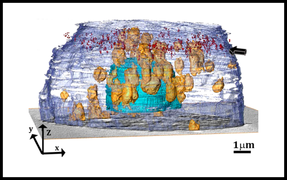
Correlative imaging of STORM and cryo soft X-ray tomography of macrophage cell incubated with acLDL. STORM measurement was preformed in RT flowing by freezing and imaging soft X-ray tomogram of the cell.
In collaboration with the group of Prof. Lia Addadi (Weizmann Institute of Science)