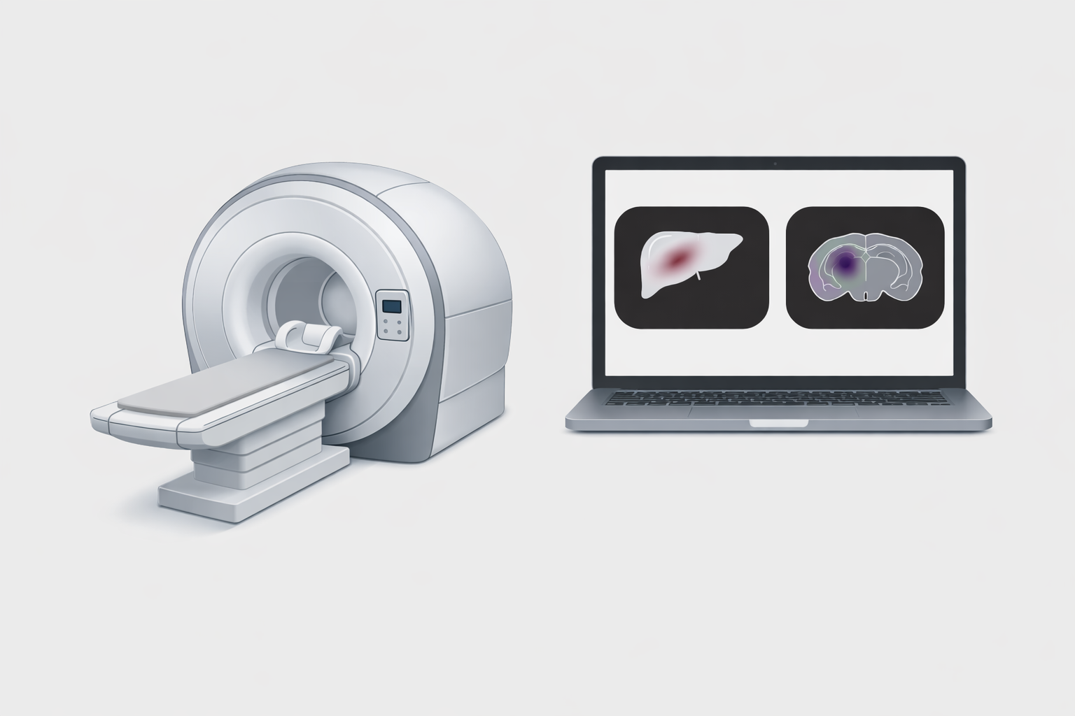
We apply synthetic chemistry, genetic engineering, advanced MRI approaches, and high-end imaging techniques to create a molecular imaging toolbox that enables longitudinal, non-invasive study of biological events in deep tissues.
Goren E., Subramani B., Avram L., Falkovich A. H., Perlman O. & Bar-Shir A.
(2025)
Journal of the American Chemical Society.
147,
22,
p. 18972-18981
The reliance of modern technology growth on lanthanides presents dual challenges: securing sustainable sources from natural or recycled materials and reducing environmental harm from waste discharge. However, the similar ionic radii, oxidation states, and binding affinities of Ln3+ ions hinder their nondestructive detection in mixtures. Furthermore, the overlap of spectroscopic signals and the inapplicability for opaque solutions limit the harness of luminescent sensors for differentiating one Ln3+ from another. Here, we introduce 19F-paramagnetic guest exchange saturation transfer magnetic resonance fingerprinting (19F-paraGEST MRF), a rapid signal acquisition, encoding, and analysis approach for detecting specific Ln3+ in mixtures. Based on a small-sized experimental 19F-paraGEST data set, we generated a de novo dictionary of ∼2500 combinations of Ln3+ mixtures, resulting in ∼7,000,000 simulated 19F-paraGEST MRF patterns of different Ln3+ concentrations. This dictionary was later used for computational pattern recognition of experimental NMR signal evolutions (\u201cfingerprints\u201d), utilizing a rapid computational approach executable on a standard laptop within seconds. Hence, fast and reliable multiplexed lanthanide detection in complex mixtures was enabled. Demonstrated through the analysis of lanthanides content of permanent magnets from a hard disk drive, this MR-based method paves the way for broader applications of lanthanide detection in murky, nontransparent mixtures and further exploration of supramolecular sensors in diverse scenarios.
Allouche-Arnon H., Montrazi E. T., Subramani B., Fisler M., Spigel I., Frydman L., Mehlman T., Brandis A., Harris T. & Bar-Shir A.
(2024)
Journal of the American Chemical Society.
146,
46,
p. 31624-31632
The ability to obtain quantitative spatial information on subcellular processes of deep tissues in vivo has been a long-standing challenge for molecular magnetic resonance imaging (MRI) approaches. This challenge remains even more so for quantifying readouts of genetically engineered MRI reporters. Here, we set to overcome this challenge with a molecular system designed to obtain quantitative 2H-MRI maps of a gene reporter. To this end, we synthesized deuterated thymidine, d3-thy, with three magnetically equivalent deuterons at its methyl group (-CD3), showing a singlet peak with a characteristic 2H-NMR frequency (δ = 1.7 ppm). The upfield 3.0 ppm offset from the chemical shift of the HDO signal (δ = 4.7 ppm) allows for spectrally resolving the two 2H NMR signals and quantifying the concentration of d3-thy based on the known concentration of a tissues HDO. Following systemic administration of d3-thy, its accumulation as d3-thy monophosphate in cells expressing the human thymidine kinase 1 (hTK1) transgene was mapped with 2H-MRI. The data obtained in vivo show the ability to use the d3-thy/hTK1 pair as a reporter probe/reporter gene system to quantitatively map transgene expression with MRI. Relying on a structurally unmodified reporter probe (d3-thy) to image the expression of unmutated human protein (hTK1) shows the potential of molecular imaging with 2H-MRI to monitor gene reporters and other relevant biological targets.
Galisova A., Zahradnik J., Allouche-Arnon H., Morandi M. I., Abou Karam P., Fisler M., Avinoam O., Regev-Rudzki N., Schreiber G. & Bar-Shir A.
(2022)
ACS Nano.
16,
8,
p. 12276-12289
The elucidation of viral-receptor interactions and an understanding of virus-spreading mechanisms are of great importance, particularly in the era of a pandemic. Indeed, advances in computational chemistry, synthetic biology, and protein engineering have allowed precise prediction and characterization of such interactions. Nevertheless, the hazards of the infectiousness of viruses, their rapid mutagenesis, and the need to study viral-receptor interactions in a complex in vivo setup call for further developments. Here, we show the development of biocompatible genetically engineered extracellular vesicles (EVs) that display the receptor binding domain (RBD) of SARS-CoV-2 on their surface as coronavirus mimetics (EVsRBD). Loading EVsRBD with iron oxide nanoparticles makes them MRI-visible and, thus, allows mapping of the binding of RBD to ACE2 receptors noninvasively in live subjects. Moreover, we show that EVsRBD can be modified to display mutants of the RBD of SARS-CoV-2, allowing rapid screening of currently raised or predicted variants of the virus. The proposed platform thus shows relevance and cruciality in the examination of quickly evolving pathogenic viruses in an adjustable, fast, and safe manner. Relying on MRI for visualization, the presented approach could be considered in the future to map ligand-receptor binding events in deep tissues, which are not accessible to luminescence-based imaging.
Tirukoti N. D., Avram L., Haris T., Lerner B., Diskin Posner Y., Allouche-Arnon H. & Bar-Shir A.
(2021)
Journal of the American Chemical Society.
143,
30,
p. 11751-11758
Fast ion-chelate dissociation rates and weak ion-chelate affinities are desired kinetic and thermodynamic features for imaging probes to allow reversible binding and to prevent deviation from basal ionic levels. Nevertheless, such properties often result in poor readouts upon ion binding, frequently result in low ion specificity, and do not allow the detection of a wide range of concentrations. Herein, we show the design, synthesis, characterization, and implementation of a Zn2+-probe developed for MRI that possesses reversible Zn2+-binding properties with a rapid dissociation rate (koff = 845 ± 35 s1) for the detection of a wide range of biologically relevant concentrations. Benefiting from the implementation of chemical exchange saturation transfer (CEST), which is here applied in the 19F-MRI framework in an approach termed ion CEST (iCEST), we demonstrate the ability to map labile Zn2+ with spectrally resolved specificity and with no interference from competitive cations. Relying on fast koff rates for enhanced signal amplification, the use of iCEST allowed the designed fluorinated chelate to experience weak Zn2+-binding affinity (Kd at the mM range), but without compromising high cationic specificity, which is demonstrated here for mapping the distribution of labile Zn2+ in the hippocampal tissue of a live mouse. This strategy for accelerating ion-chelate koff rates for the enhancement of MRI signal amplifications without affecting ion specificity could open new avenues for the design of additional probes for other metal ions beyond zinc.