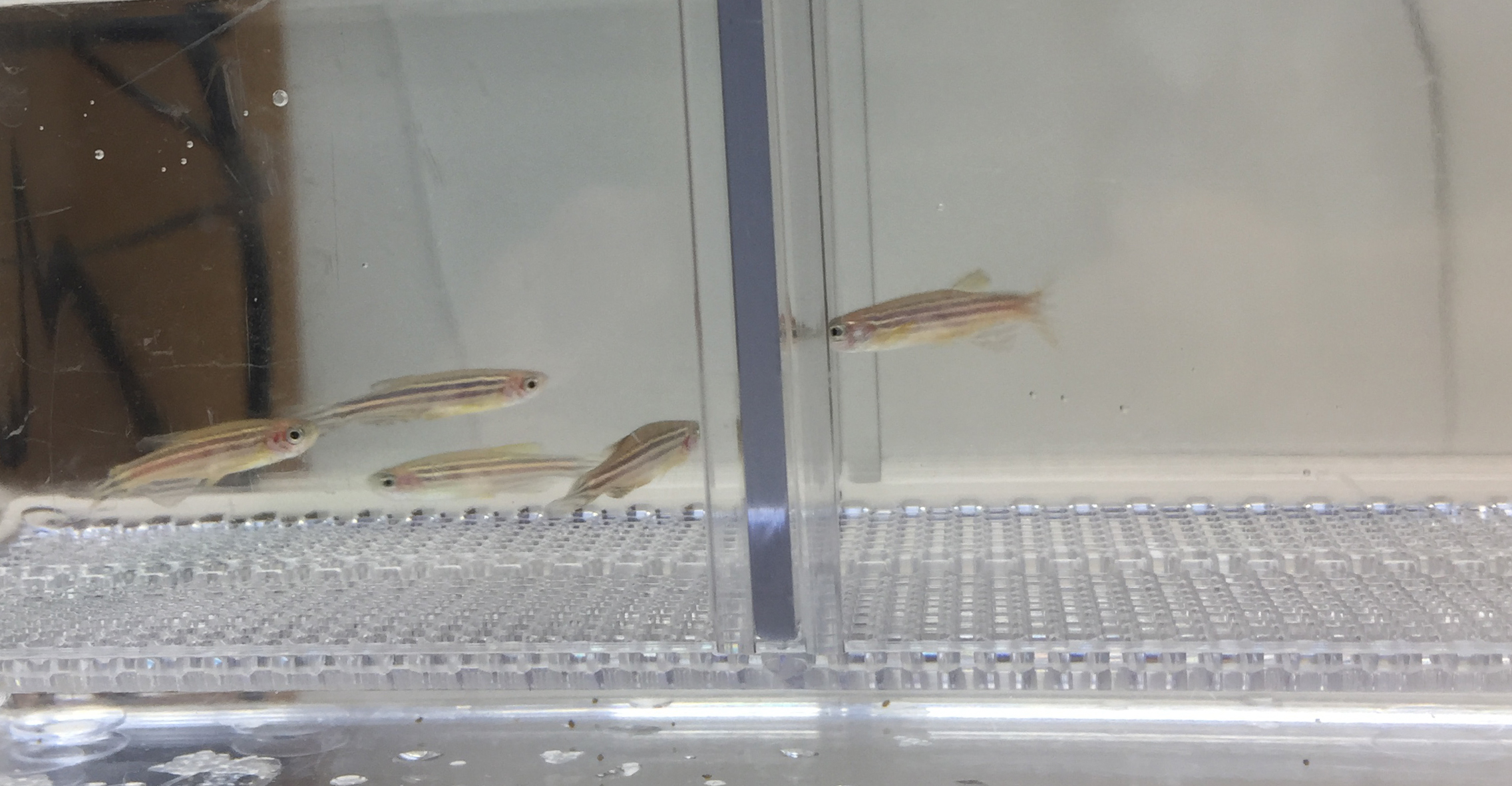
[Skip Header and Navigation] [Jump to Main Content]
Weizmann Institute of Science
Home
Department of Molecular Cell Biology

  • Home
  • Research
  • Publications
  • People
  • Photo Gallery
  • Videos
  • In the News
  • Positions
  • Links

  • Members Only

Research

The hypothalamus regulates responses to physical and psychological stress by integrating sensory information from virtually the entire body and in turn affecting endocrine, metabolic and behavioral activities in order to restore homeostasis.  Abnormalities in the development of the hypothalamus have been associated with neurological conditions such as depression, chronic stress, autism and obesity.  If we are to understand the genetic causes of such neuro-developmental disorders, we must first gain an understanding of hypothalamic development.  However, uncovering the developmental processes that govern formation of the hypothalamus has proved challenging due to the immense number of distinct neuronal cell types that arise in very close proximity to one another and connect with many targets throughout the nervous system. It is unclear how the diversity of hypothalamic cell fates is coordinated so precisely, achieving very accurate numbers, types and positions of neurons.

We chose to study hypothalamic neural specification in zebrafish, a vertebrate organism readily amenable to genetic manipulations, and whose embryos are optically transparent, allowing in vivo analysis of both hypothalamic neurons and their circuits. We employ genetic approaches in combination with state-of-the art imaging techniques to identify critical determinants of hypothalamic differentiation and function (Machluf et al. 2011).  Specifically, we have been addressing several key questions relating to the patterning, specification and morphogenesis of the hypothalamus. Finally, we have recently begun studying activity-dependent gene regulation in response to homeostatic challenges to the mature hypothalamus.


Origins of early hypothalamic precursors

The hypothalamus is a major part of the basal diencephalon.  To better understand the embryonic development and morphogenesis of the diencephalon we employed two-photon microscopy to photo-activate a fluorescent dye in a living zebrafish embryo (Russek-Blum et al. 2010).  This approach facilitated precise labeling of defined neural progenitors in the prospective diencephalon of the zebrafish neural plate and allowed us to create a fate map of the zebrafish diencephalon at a higher spatial resolution than previously described (Russek-Blum et al. 2008, 2009).  


The genetic requirements for the establishment of hypothalamic fates

Our fate mapping analyses showed that progenitors of specific hypothalamic cell-types originate from stereotypical positions in the neural plate suggesting that their identity is intrinsically encoded by a specific set of molecules. Indeed, we have identified a molecular transcriptional code underlying different stages in hypothalamic differentiation and showed that hypothalamic specification requires the sequential activity of pan-neuronal and type-specific transcription factors (Borodovsky et al. 2009).  This study provided new evidence linking early neurogenesis with a specific differentiation program.  

We also address the mechanism by which coordinated generation of various hypothalamic neurons is achieved, by focusing on zebrafish dopaminergic and oxytocinergic neurons representing diverse hypothalamic cell types.  Interestingly, we revealed that quantitative differences in a single intrinsic factor– Otp leads to specific differentiation programs that promote spatio-temporally distinct dopaminergic and oxytocinergic identities (Blechman et al. 2007).


Determining the size of hypothalamic neuronal population

The generation and maintenance of hypothalamic neurons must be tightly coordinated for the brain to function properly. We took advantage of the small cell population size in the zebrafish to quantify the number of individual neuronal cell types (Russek-Blum et al. 2008).  We learned that individual hypothalamic clusters reach an almost invariant cell number during development. The question is which developmental cues regulate the nearly fixed number of hypothalamic neuronal types and how? Surprisingly, we found that some elements of cell fate determination occur together with early patterning of the nervous system.  In this regards, we showed that the secreted patterning protein Wnt plays a critical role in delineating the upper and lower size limits of selected hypothalamic neuronal clusters (Russek-Blum et al. 2008).


Morphogenesis of the hypothalamic neuro-vascular interface

The hypothalamo-neurohypophyseal system (HNS) is a main conduit by which the brain exerts control over peripheral organs.  The HNS neuropeptides, oxytocin and arginine-vasopressin, are transported along axons and secreted onto fenestrated capillaries in the neurohypophysis, where they enter the general circulation and affect peripheral tissues such as the kidneys and the uterus (Gutnick and Levkowitz 2012).  Although the interface between the neurons and blood vessels of the HNS is essential for direct transfer of neuropeptides from the central nervous system to the blood, its formation during embryonic development has yet to be understood.

We have generated a transgenic zebrafish line, in which oxytocinergic neurons, their axonal termini and the neurohypophyseal blood vessels are labeled (Blechman et al. 2011, Gutnick et al. 2011).  This offers a unique tool to study the development and function of the HNS in vivo without the need for surgical intervention. We are studying the signaling events that control the formation of the axonal-vascular interactions during neurohypophyseal development.  For example, we revealed that secretion of the neuropeptide oxytocin from HNS axons serves as a localized cue that mediates formation a tight neuro-vascular interface  ( Gutnick et al. 2011).


Role of ‘developmental’ factors in physiological hypothalamic function

We noticed that the expression of some genetic determinant of hypothalamic development is maintained in the adult fish and mouse suggesting that they may actually play an active role in the physiological functions of the adult hypothalamus.  To examine this possibility we are currently employing several established and novel stress paradigms in both mice and fish in order to uncover the transcriptional response to homeostatic challenges.  We use Chromatin Immunoprecipitation of developmetal transcription factors in search of new central mediators of stress challenge. This analysis has already yielded several target genes previously found to be genetically linked to psychiatric and neurodevelopmental disorders as well as a number of novel candidate mediators of stress (Amir-Zilberstein et al. 2012).  This project promises to shed new light on the molecular mechanisms linking neuro-developmental factors to neurological disorders associated with hypothalamic dysfunction.

[Jump to Top] [Jump to Main Content]