Publications
2023
-
(2023) EMBO Reports. e56114. Abstract[All authors]
Vesicular transport is a means of communication. While cells can communicate with each other via secretion of extracellular vesicles, less is known regarding organelle-to organelle communication, particularly in the case of mitochondria. Mitochondria are responsible for the production of energy and for essential metabolic pathways in the cell, as well as fundamental processes such as apoptosis and aging. Here, we show that functional mitochondria isolated from Saccharomyces cerevisiae release vesicles, independent of the fission machinery. We isolate these mitochondrial-derived vesicles (MDVs) and find that they are relatively uniform in size, of about 100 nm, and carry selective protein cargo enriched for ATP synthase subunits. Remarkably, we further find that these MDVs harbor a functional ATP synthase complex. We demonstrate that these vesicles have a membrane potential, produce ATP, and seem to fuse with naive mitochondria. Our findings reveal a possible delivery mechanism of ATP-producing vesicles, which can potentially regenerate ATP-deficient mitochondria and may participate in organelle-to-organelle communication.
-
(2023) Parasites & vectors. 16, 1, p. - 14. Abstract[All authors]
Background
Plasmodium falciparum (Pf) is the leading protozoan causing malaria, the most devastating parasitic disease. To ensure transmission, a small subset of Pf parasites differentiate into the sexual forms (gametocytes). Since the abundance of these essential parasitic forms is extremely low within the human host, little is currently known about the molecular regulation of their sexual differentiation, highlighting the need to develop tools to investigate Pf gene expression during this fundamental mechanism.Methods
We developed a high-throughput quantitative Reverse-Transcription PCR (RT-qPCR) platform to robustly monitor Pf transcriptional patterns, in particular, systematically profiling the transcriptional pattern of a large panel of gametocyte-related genes (GRG). Initially, we evaluated the technical performance of the systematic RT-qPCR platform to ensure it complies with the accepted quality standards for: (i) RNA extraction, (ii) cDNA synthesis and (iii) evaluation of gene expression through RT-qPCR. We then used this approach to monitor alterations in gene expression of a panel of GRG upon treatment with gametocytogenesis regulators.Results
We thoroughly elucidated GRG expression profiles under treatment with the antimalarial drug dihydroartemisinin (DHA) or the metabolite choline over the course of a Pf blood cycle (48 h). We demonstrate that both significantly alter the expression pattern of PfAP2-G, the gametocytogenesis master regulator. However, they also markedly modify the developmental rate of the parasites and thus might bias the mRNA expression. Additionally, we screened the effect of the metabolites lactate and kynurenic acid, abundant in severe malaria, as potential regulators of gametocytogenesis.Conclusions
Our data demonstrate that the high-throughput RT-qPCR method enables studying the immediate transcriptional response initiating gametocytogenesis of the parasites from a very low volume of malaria-infected RBC samples. The obtained data expand the current knowledge of the initial alterations in mRNA profiles of GRG upon treatment with reported regulators. In addition, using this method emphasizes that asexual parasite stage composition is a crucial element that must be considered when interpreting changes in GRG expression by RT-qPCR, specifically when screening for novel compounds that could regulate Pf sexual differentiation.
2022
-
(2022) PNAS Nexus. 1, 4, p. 1-11 Abstract[All authors]
Extracellular vesicles (EVs) transfer bioactive molecules between cells in a process reminiscent of enveloped viruses. EV cargo delivery is thought to occur by protein-mediated and pH-dependent membrane fusion of the EV and the cellular membrane. However, there is a lack of methods to identify the fusion proteins and resolve their mechanism. We developed and benchmarked an in vitro biophysical assay to investigate EV membrane fusion. The assay was standardized by directly comparing EV and viral fusion with liposomes. We show that EVs and retroviruses fuse with liposomes mimicking the membrane composition of the late endosome in a pH-and protein-dependent manner. Moreover, we directly visualize the stages of membrane fusion using cryo-electron tomography. We find that, unlike most retroviruses, EVs remain fusogenic after acidification and reneutralization. These results provide novel insights into the EV cargo delivery mechanism and an experimental approach to identify the EV fusion machinery. Significance Statement: Extracellular vesicles (EVs) are membrane-bound compartments containing bioactive cargos, released from cells to mediate inter-cellular communication. EV cargo delivery is thought to occur via EV-to-cell membrane fusion. However, EV fusion has not been directly visualized and the fusion mechanism remains uncharacterized. We benchmark an in vitro assay that recapitulates EVs fusion with liposomes using enveloped viruses as control. We show that EV-liposome fusion recapitulates features previously reported for EV cargo delivery, and that the fusion process can be visualized at a nanometric resolution using cryo-electron mi-croscopy. Moreover, our data suggest that unlike most enveloped viruses, EV fusion is not inactivated upon exposure to acidic pH in the absence of target membranes, which is consistent with biogenesis in multivesicular bodies.
-
(2022) ACS Nano. 16, 8, Abstract[All authors]
The elucidation of viral-receptor interactions and an understanding of virus-spreading mechanisms are of great importance, particularly in the era of a pandemic. Indeed, advances in computational chemistry, synthetic biology, and protein engineering have allowed precise prediction and characterization of such interactions. Nevertheless, the hazards of the infectiousness of viruses, their rapid mutagenesis, and the need to study viral-receptor interactions in a complex in vivo setup call for further developments. Here, we show the development of biocompatible genetically engineered extracellular vesicles (EVs) that display the receptor binding domain (RBD) of SARS-CoV-2 on their surface as coronavirus mimetics (EVsRBD). Loading EVsRBD with iron oxide nanoparticles makes them MRI-visible and, thus, allows mapping of the binding of RBD to ACE2 receptors noninvasively in live subjects. Moreover, we show that EVsRBD can be modified to display mutants of the RBD of SARS-CoV-2, allowing rapid screening of currently raised or predicted variants of the virus. The proposed platform thus shows relevance and cruciality in the examination of quickly evolving pathogenic viruses in an adjustable, fast, and safe manner. Relying on MRI for visualization, the presented approach could be considered in the future to map ligand-receptor binding events in deep tissues, which are not accessible to luminescence-based imaging.
-
(2022) Malaria Immunology. p. 133-145 Abstract
Malaria is one the most devastating infectious diseases in the world: of the five malaria-associated parasites, Plasmodium falciparum and P. vivax are the most pathogenic and widespread, respectively. P. falciparum invades human red blood cells (RBCs), releasing extracellular vesicles (Pf-EV) carrying DNA, RNA and protein cargo components involved in host-pathogen communications in the course of the disease. Different strategies have been used to analyze Pf-EV biophysically and chemically. Atomic force microscopy (AFM) stands out as a powerful tool for rendering high quality images of extracellular vesicles. In this technique, a sharp tip attached to a cantilever reconstructs the topographic surface of the extracellular vesicles and probes their nano-mechanical properties based on force–distance curves. Here, we describe a method to separate Pf-EV using differential ultracentrifugation, followed by nanoparticle tracking analysis (NTA) to quantify and estimate the size distribution. Finally, the AFM imaging procedure on Pf-EV adsorbed on a Mg2+-modified mica surface is detailed.
-
(2022) EMBO Reports. e54755. Abstract[All authors]
Malaria is the most serious mosquito-borne parasitic disease, caused mainly by the intracellular parasite Plasmodium falciparum. The parasite invades human red blood cells and releases extracellular vesicles (EVs) to alter its host responses. It becomes clear that EVs are generally composed of sub-populations. Seeking to identify EV subpopulations, we subject malaria-derived EVs to size-separation analysis, using asymmetric flow field-flow fractionation. Multi-technique analysis reveals surprising characteristics: we identify two distinct EV subpopulations differing in size and protein content. Small EVs are enriched in complement-system proteins and large EVs in proteasome subunits. We then measure the membrane fusion abilities of each subpopulation with three types of host cellular membranes: plasma, late and early endosome. Remarkably, small EVs fuse to early endosome liposomes at significantly greater levels than large EVs. Atomic force microscope imaging combined with machine-learning methods further emphasizes the difference in biophysical properties between the two subpopulations. These results shed light on the sophisticated mechanism by which malaria parasites utilize EV subpopulations as a communication tool to target different cellular destinations or host systems.
-
(2022) Faraday Discussions. 239, Abstract
Malaria is a potentially fatal infectious disease caused by the obligate intracellular parasite Plasmodium falciparum. The parasite infects human red blood cells (RBC) and derives nutrition by catabolism of hemoglobin. As amino acids are assimilated from the protein component, the toxic heme is released. Molecular heme is detoxified by rapid sequestration to physiologically insoluble hemozoin crystals within the parasite's digestive vacuole (DV). Common antimalarial drugs interfere with this crystallization process, leaving the parasites vulnerable to the by-product of their own metabolism. A fundamental debate with important implications on drug mechanism regards the chemical environment of crystallization in situ, whether aqueous or lipid. This issue had been addressed previously by cryogenic soft X-ray tomography. We employ cryo-scanning transmission electron tomography (CSTET) to probe parasite cells throughout the life cycle in a fully hydrated, vitrified state at higher resolution. During the acquisition of CSTET data, Bragg diffraction from the hemozoin provides a uniquely clear view of the crystal boundary at nanometer resolution. No intermediate medium, such as a lipid coating or shroud, could be detected surrounding the crystals. The present study describes a unique application of CSTET in the study of malaria. The findings can be extended to evaluate new drug candidates affecting hemozoin crystal growth.
-
(2022) Journal of Extracellular Biology. 1, 2, Abstract[All authors]
Glycoconjugates on extracellular vesicles (EVs) play a vital role in internalization and mediate interaction as well as regulation of the host immune system by viruses, bacteria, and parasites. During their intraerythrocytic life‐cycle stages, malaria parasites, Plasmodium falciparum (Pf) mediate the secretion of EVs by infected red blood cells (RBCs) that carry a diverse range of parasitic and host‐derived molecules. These molecules facilitate parasite‐parasite and parasite‐host interactions to ensure parasite survival.
To date, the number of identified Pf genes associated with glycan synthesis and the repertoire of expressed glycoconjugates is relatively low. Moreover, the role of Pf glycans in pathogenesis is mostly unclear and poorly understood. As a result, the expression of glycoconjugates on Pf‐derived EVs or their involvement in the parasite life‐cycle has yet to be reported.
Herein, we show that EVs secreted by Pf‐infected RBCs carry significantly higher sialylated complex N‐glycans than EVs derived from healthy RBCs. Furthermore, we reveal that EV uptake by host monocytes depends on N‐glycoproteins and demonstrate that terminal sialic acid on the N‐glycans is essential for uptake by human monocytes. Our results provide the first evidence that Pf exploits host sialylated N‐glycans to mediate EV uptake by the human immune system cells. -
(2022) Frontiers in Cellular and Infection Microbiology. 11, 739628. Abstract
Extracellular vesicles (EVs) are produced by across almost all the living kingdoms and play a crucial role in cell-cell communication processes. EVs are especially important for pathogens, as Plasmodium falciparum (Pf) parasite, the leading causing species in human malaria. Malaria parasites are able to modulate the host immune response from a distance via delivering diverse cargo components inside the EVs, such as proteins and nucleic acids. We have previously shown that imaging flow cytometry (IFC) can be effectively used to monitor the uptake of different cargo components of malaria derived EVs by host human monocytes. Here, we take this approach one step further and demonstrate that we can directly investigate the dynamics of the cargo distribution pattern over time by monitoring its distribution within two different recipient cells of the immune system, monocytes vs macrophages. By staining the RNA cargo of the vesicles and monitor the signal we were able to evaluate the kinetics of its delivery and measure different parameters of the cargo’s distribution post internalization. Interestingly, we found that while the level of the EV uptake is similar, the pattern of the signal for RNA cargo distribution is significantly different between these two recipient immune cells. Our results demonstrate that this method can be applied to study the distribution dynamics of the vesicle cargo post uptake to different types of cells. This can benefit significantly to our understanding of the fate of cargo components post vesicle internalization in the complex interface between pathogen-derived vesicles and their host recipient cells.
2021
-
(2021) Cell Death & Disease. 12, 11, p. 1059-1059 Abstract[All authors]
AbstractNecroptosis is a regulated and inflammatory form of cell death. We, and others, have previously reported that necroptotic cells release extracellular vesicles (EVs). We have found that necroptotic EVs are loaded with proteins, including the phosphorylated form of the key necroptosis-executing factor, mixed lineage kinase domain-like kinase (MLKL). However, neither the exact protein composition, nor the impact, of necroptotic EVs have been delineated. To characterize their content, EVs from necroptotic and untreated U937 cells were isolated and analyzed by mass spectrometry-based proteomics. A total of 3337 proteins were identified, sharing a high degree of similarity with exosome proteome databases, and clearly distinguishing necroptotic and control EVs. A total of 352 proteins were significantly upregulated in the necroptotic EVs. Among these were MLKL and caspase-8, as validated by immunoblot. Components of the ESCRTIII machinery and inflammatory signaling were also upregulated in the necroptotic EVs, as well as currently unreported components of vesicle formation and transport, and necroptotic signaling pathways. Moreover, we found that necroptotic EVs can be phagocytosed by macrophages to modulate cytokine and chemokine secretion. Finally, we uncovered that necroptotic EVs contain tumor neoantigens, and are enriched with components of antigen processing and presentation. In summary, our study reveals a new layer of regulation during the early stage of necroptosis, mediated by the secretion of specific EVs that influences the microenvironment and may instigate innate and adaptive immune responses. This study sheds light on new potential players in necroptotic signaling and its related EVs, and uncovers the functional tasks accomplished by the cargo of these necroptotic EVs.
-
(2021) Nature Communications. 12, 4851. Abstract[All authors]
The chemokine CXCL10 is associated with pathogenesis of cerebral malaria in Plasmodium falciparum infection. Here the authors show that P. falciparum produces extracellular vesicles laden with RNAs that are taken up by monocytes resulting in a RIG-I and HUR-1 mediated mechanism of inhibition of CXCL10 protein translation.
-
(2021) Beilstein Journal of Nanotechnology. 12, p. 878-901 Abstract
Progress in computing capabilities has enhanced science in many ways. In recent years, various branches of machine learning have been the key facilitators in forging new paths, ranging from categorizing big data to instrumental control, from materials design through image analysis. Deep learning has the ability to identify abstract characteristics embedded within a data set, subsequently using that association to categorize, identify, and isolate subsets of the data. Scanning probe microscopy measures multimodal surface properties, combining morphology with electronic, mechanical, and other characteristics. In this review, we focus on a subset of deep learning algorithms, that is, convolutional neural networks, and how it is transforming the acquisition and analysis of scanning probe data.
-
-
-
(2021) Circulation (New York, N.Y.). 143, 25, p. 2475-2493 Abstract[All authors]
Background: The role of epicardial fat (eFat)-derived extracellular vesicles (EVs) in the pathogenesis of atrial fibrillation (AF) has never been studied. We tested the hypothesis that eFat-EVs transmit proinflammatory, profibrotic, and proarrhythmic molecules that induce atrial myopathy and fibrillation. Methods: We collected eFat specimens from patients with (n=32) and without AF (n=30) during elective heart surgery. eFat samples were grown as organ cultures, and the culture medium was collected every 2 days. We then isolated and purified eFat-EVs from the culture medium, and analyzed the EV number, size, morphology, specific markers, encapsulated cytokines, proteome, and microRNAs. Next, we evaluated the biological effects of unpurified and purified EVs on atrial mesenchymal stromal cells and endothelial cells in vitro. To establish a causal association between eFat-EVs and vulnerability to AF, we modeled AF in vitro using induced pluripotent stem cell–derived cardiomyocytes. Results: Microscopic examination revealed excessive inflammation, fibrosis, and apoptosis in fresh and cultured eFat tissues. Cultured explants from patients with AF secreted more EVs and harbored greater amounts of proinflammatory and profibrotic cytokines, and profibrotic microRNA, as well, than those without AF. The proteomic analysis confirmed the distinctive profile of purified eFat-EVs from patients with AF. In vitro, purified and unpurified eFat-EVs from patients with AF had a greater effect on proliferation and migration of human mesenchymal stromal cells and endothelial cells, compared with eFat-EVs from patients without AF. Last, whereas eFat-EVs from patients with and without AF shortened the action potential duration of induced pluripotent stem cell–derived cardiomyocytes, only eFat-EVs from patients with AF induced sustained reentry (rotor) in induced pluripotent stem cell–derived cardiomyocytes. Conclusions: We show, for the first time, a distinctive proinflammatory, profibrotic, and proarrhythmic signature of eFat-EVs from patients with AF. Our findings uncover another pathway by which eFat promotes the development of atrial myopathy and fibrillation.
-
(2021) Cancer research (Chicago, Ill.). 81, 7, p. 1639-1653 Abstract[All authors]
Gastric cancer is the 3rd most lethal cancer worldwide, and evaluation of the genomic status of gastric cancer cells has not translated into effective prognostic or therapeutic strategies. We therefore hypothesize that outcomes may depend on the tumor microenvironment (TME), in particular, cancer-associated fibroblasts (CAF). However, very little is known about the role of CAFs in gastric cancer. To address this, we mapped the transcriptional landscape of human gastric cancer stroma by microdissection and RNA sequencing of CAFs from gastric cancer patients. A stromal gene signature was associated with poor disease outcome, and the transcription factor heat shock factor 1 (HSF1) regulated the signature. HSF1 upregulated inhibin subunit beta A (INHBA) and thrombospondin 2 (THBS2), which were secreted in CAF-derived extracellular vesicles (EV) to the TME to promote cancer. Together, our work provides the first transcriptional map of human gastric cancer stroma and highlights HSF1 and its transcriptional targets as potential diagnostic and therapeutic targets in the genomically stable tumor microenvironment.
-
(2021) Nature Communications. 12, 1, 1172. Abstract[All authors]
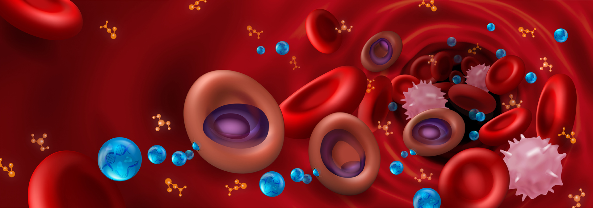
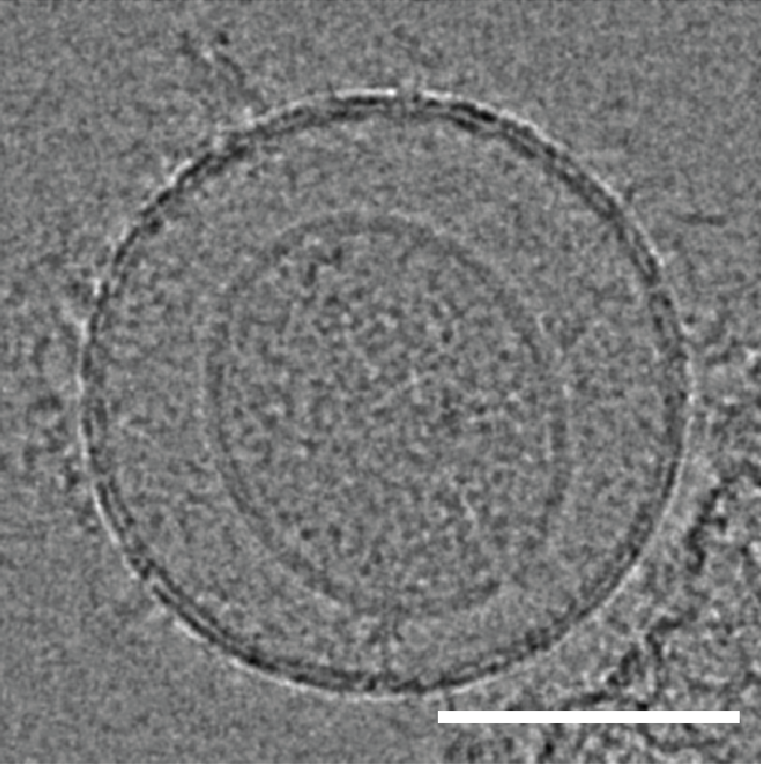
Mature red blood cells (RBCs) lack internal organelles and canonical defense mechanisms, making them both a fascinating host cell, in general, and an intriguing choice for the deadly malaria parasite Plasmodium falciparum (Pf), in particular. Pf, while growing inside its natural host, the human RBC, secretes multipurpose extracellular vesicles (EVs), yet their influence on this essential host cell remains unknown. Here we demonstrate that Pf parasites, cultured in fresh human donor blood, secrete within such EVs assembled and functional 20S proteasome complexes (EV-20S). The EV-20S proteasomes modulate the mechanical properties of naïve human RBCs by remodeling their cytoskeletal network. Furthermore, we identify four degradation targets of the secreted 20S proteasome, the phosphorylated cytoskeletal proteins β-adducin, ankyrin-1, dematin and Epb4.1. Overall, our findings reveal a previously unknown 20S proteasome secretion mechanism employed by the human malaria parasite, which primes RBCs for parasite invasion by altering membrane stiffness, to facilitate malaria parasite growth.
2020
-
(2020) Frontiers in Cellular and Infection Microbiology. 10, 380. Abstract
Leishmania infection causes considerable human morbidity and may develop into a deadly visceral form in endemic regions. The parasite infects macrophages where they can replicate intracellularly. Furthermore, they modulate host immune responses by using virulence factors (lipophosphoglycan, glycoprotein-63, and others) that promote survival inside the cells. Extracellular vesicles (EVs) released by parasites are important for cell-cell communication in the proinflammatory milieu modulating the establishment of infection. However, information on the ability of EVs from different Leishmania species to modulate inflammatory responses is scarce, especially from those species causing different clinical manifestations (visceral vs. cutaneous). The purpose of this study was to compare macrophage activation using EVs from three Leishmania species from New World including L. infantum, L. braziliensis, and L. amazonensis. EVs were released from promastigote forms, purified by ultracentrifugation and quantitated by Nanoparticle Tracking Analysis (NTA) prior to murine macrophage exposure. NTA analysis did not show any differences in the EV sizes among the strains. EVs from L. braziliensis and L. infantum failed to induce a pro-inflammatory response. EVs from both L. infantum WT and LPG-deficient mutant (LPG-KO) did not show any differences in their interaction with macrophages, suggesting that LPG solely was not determinant for activation. On the other hand, EVs from L. amazonensis were immunomodulatory inducing NO, TNF-alpha, IL-6, and IL-10 via TLR4 and TLR2. To determine whether such activation was related to NF-kappa B p65 translocation, THP-1 macrophage cells were exposed to EVs. In the same way, only EVs from L. amazonensis exhibited a highly percentage of cells positive for NF-kappa B. Our results suggest an important role of EVs in determining the pattern of immune response depending on the parasite species. For L. infantum, LPG was not determinant for the activation.
-
(2020) Biomedicines. 8, 5, 98. Abstract
Extracellular vesicles (EVs) are cell-derived membrane-bound structures that are believed to play a major role in intercellular communication by allowing cells to exchange proteins and genetic cargo between them. In particular, pathogens, such as the malaria parasite Plasmodium (P.) falciparum, utilize EVs to promote their growth and to alter their host's response. Thus, better characterization of these secreted organelles will enhance our understanding of the cellular processes that govern EVs' biology and pathological functions. Here we present a method that utilizes a high-end flow cytometer system to characterize small EVs, i.e., with a diameter less than 200 nm. Using this method, we could evaluate different parasite-derived EV populations according to their distinct cargo by using antibody-free labeling. It further allows to closely monitor a sub-population of vesicles carrying parasitic DNA cargo. This ability paves the way to conducting a more 'educated' analysis of the various EV cargo components.
-
(2020) Frontiers in Cellular and Infection Microbiology. 10, 99. Abstract[All authors]
Extracellular vesicles (EVs) shed by trypomastigote forms of Trypanosoma cruzi have the ability to interact with host tissues, increase invasion, and modulate the host innate response. In this study, EVs shed from T. cruzi or T.cruzi-infected macrophages were investigated as immunomodulatory agents during the initial steps of infection. Initially, by scanning electron microscopy and nanoparticle tracking analysis, we determined that T. cruzi-infected macrophages release higher numbers of EVs (50-300 nm) as compared to non-infected cells. Using Toll-like-receptor 2 (TLR2)-transfected CHO cells, we observed that pre-incubation of these host cells with parasite-derived EVs led to an increase in the percentage of infected cells. In addition, EVs from parasite or T.cruzi-infected macrophages or not were able to elicit translocation of NF-kappa B by interacting with TLR2, and as a consequence, to alter the EVs the gene expression of proinflammatory cytokines (TNF-alpha, IL-6, and IL-1 beta), and STAT-1 and STAT-3 signaling pathways. By proteomic analysis, we observed highly significant changes in the protein composition between non-infected and infected host cell-derived EVs. Thus, we observed the potential of EVs derived from T. cruzi during infection to maintain the inflammatory response in the host.
-
(2020) EMBO Reports. 21, 1, 47882. Abstract[All authors]
During the chronic stage of Schistosoma infection, the female lays fertile eggs, triggering a strong anti-parasitic type 2 helper T-cell (Th2) immune response. It is unclear how this Th2 response gradually declines even though the worms live for years and continue to produce eggs. Here, we show that Schistosoma mansoni downregulates Th2 differentiation in an antigen-presenting cell-independent manner, by modulating the Th2-specific transcriptional program. Adult schistosomes secrete miRNA-harboring extracellular vesicles that are internalized by Th cells in vitro. Schistosomal miRNAs are found also in T helper cells isolated from Peyer's patches and mesenteric lymph nodes of infected mice. In T helper cells, the schistosomal miR-10 targets MAP3K7 and consequently downmodulates NF-kappa B activity, a critical transcription factor for Th2 differentiation and function. Our results explain, at least partially, how schistosomes tune down the Th2 response, and provide further insight into the reciprocal geographic distribution between high prevalence of parasitic infections and immune disorders such as allergy. Furthermore, this worm-host crosstalk mechanism can be harnessed to develop diagnostic and therapeutic approaches for human schistosomiasis and Th2-associated diseases.
-
(2020) Proteins-Structure Function And Bioinformatics. 88, 1, p. 187-195 Abstract
Many human pathogens use host cell-surface receptors to attach and invade cells. Often, the host-pathogen interaction affinity is low, presenting opportunities to block invasion using a soluble, high-affinity mimic of the host protein. The Plasmodium falciparum reticulocyte-binding protein homolog 5 (RH5) provides an exciting candidate for mimicry: it is highly conserved and its moderate affinity binding to the human receptor basigin (K
D ≥1 μM) is an essential step in erythrocyte invasion by this malaria parasite. We used deep mutational scanning of a soluble fragment of human basigin to systematically characterize point mutations that enhance basigin affinity for RH5 and then used Rosetta to design a variant within the sequence space of affinity-enhancing mutations. The resulting seven-mutation design exhibited 1900-fold higher affinity (K
D approximately 1 nM) for RH5 with a very slow binding off rate (0.23 h
−1) and reduced the effective Plasmodium growth-inhibitory concentration by at least 10-fold compared to human basigin. The design provides a favorable starting point for engineering on-rate improvements that are likely to be essential to reach therapeutically effective growth inhibition.
2019
-
(2019) Cellular Microbiology. 21, 7, 13021. Abstract
Protozoan pathogens secrete nanosized particles called extracellular vesicles (EVs) to facilitate their survival and chronic infection. Here, we show the inhibition by Plasmodium berghei NK65 blood stage-derived EVs of the proliferative response of CD4(+) T cells in response to antigen presentation. Importantly, these results were confirmed in vivo by the capacity of EVs to diminish the ovalbumin-specific delayed type hypersensitivity response. We identified two proteins associated with EVs, the histamine releasing factor (HRF) and the elongation factor 1 alpha (EF-1 alpha) that were found to have immunosuppressive activities. Interestingly, in contrast to WT parasites, EVs from genetically HRF- and EF-1 alpha-deficient parasites failed to inhibit T cell responses in vitro and in vivo. At the level of T cells, we demonstrated that EVs from WT parasites dephosphorylate key molecules (PLC gamma 1, Akt, and ERK) of the T cell receptor signalling cascade. Remarkably, immunisation with EF-1 alpha alone or in combination with HRF conferred a long-lasting antiparasite protection and immune memory. In conclusion, we identified a new mechanism by which P. berghei-derived EVs exert their immunosuppressive functions by altering T cell responses. The identification of two highly conserved immune suppressive factors offers new conceptual strategies to overcome EV-mediated immune suppression in malaria-infected individuals.
-
(2019) Journal of Extracellular Vesicles. 8, 1, 1590116. Abstract
All living organisms secrete molecules for intercellular communication. Recent research has revealed that extracellular vesicles (EVs) play an important role in inter-organismal cell-to-cell communication by transporting diverse messenger molecules, including RNA, DNA, lipids and proteins. These discoveries have raised fundamental questions regarding EV biology. How are EVs biosynthesized and loaded with messenger/cargo molecules? How are EVs secreted into the extracellular matrix? What are the EV uptake mechanisms of recipient cells? As EVs are produced by all kind of organisms, from unicellular bacteria and protists, filamentous fungi and oomycetes, to complex multicellular life forms such as plants and animals, basic research in diverse model systems is urgently needed to shed light on the multifaceted biology of EVs and their role in inter-organismal communications. To help catalyse progress in this emerging field, a mini-symposium was held in Munich, Germany in August 2018. This report highlights recent progress and major questions being pursued across a very diverse group of model systems, all united by the question of how EVs contribute to inter-organismal communication.
-
(2019) EMBO Reports. 20, 3, 47719. Abstract
Tuberculosis remains one of the deadliest infectious diseases worldwide. Mycobacterium tuberculosis (M.tb) has developed various mechanisms to manipulate the human host, in particular by disrupting the host phagosome and the immune response. It is becoming evident that secreted extracellular vesicles (EVs) are involved in the dynamic crosstalk between M.tb and the host cells. These vesicles shuttle different cargo components, such as RNA, lipids, and proteins, between cells. In this issue of EMBO Reports, Cheng and Schorey describe a previously unknown EV-mediated process, regulating M.tb RNA loading into EVs and their internalization by naive macrophages. They identify the mycobacterial Sec2 secretion system as involved in RNA loading into EVs and show that secreted vesicles contain bacterial RNA that not only promotes IFN-beta production upon entry into target cells, but also leads to M.tb clearance via the activation of the host's RIG-I/MAVS signaling pathway. Importantly, combined treatment with secreted EVs and antibiotics decreases bacterial load in a mouse model, improving lung pathology compared to treatment with antibiotics alone.
-
-
(2019) Proteomics. 19, 1-2, 1800170. Abstract
Genetic plasticity of prokaryotic microbial communities is largely dependent on the ongoing exchange of genetic determinants by Horizontal Gene Transfer (HGT). HGT events allow beneficial genetic transitions to occur throughout microbial life, thus promoting adaptation to changing environmental conditions. Here, the significance of secreted vesicles in mediating HGT between microorganisms is discussed, while focusing on the benefits gained by vesicle-mediated gene delivery and its occurrence under different environmental cues. The potential use of secreted DNA-harboring vesicles as a mechanism of currently unresolved HGT events in eukaryotic microbes is further discussed.
-
(2019) Journal of Extracellular Vesicles. 7, 1, 1535750. Abstract[All authors]
The last decade has seen a sharp increase in the number of scientific publications describing physiological and pathological functions of extracellular vesicles (EVs), a collective term covering various subtypes of cell-released, membranous structures, called exosomes, microvesicles, microparticles, ectosomes, oncosomes, apoptotic bodies, and many other names. However, specific issues arise when working with these entities, whose size and amount often make them difficult to obtain as relatively pure preparations, and to characterize properly. The International Society for Extracellular Vesicles (ISEV) proposed Minimal Information for Studies of Extracellular Vesicles ("MISEV") guidelines for the field in 2014. We now update these "MISEV2014" guidelines based on evolution of the collective knowledge in the last four years. An important point to consider is that ascribing a specific function to EVs in general, or to subtypes of EVs, requires reporting of specific information beyond mere description of function in a crude, potentially contaminated, and heterogeneous preparation. For example, claims that exosomes are endowed with exquisite and specific activities remain difficult to support experimentally, given our still limited knowledge of their specific molecular machineries of biogenesis and release, as compared with other biophysically similar EVs. The MISEV2018 guidelines include tables and outlines of suggested protocols and steps to follow to document specific EV-associated functional activities. Finally, a checklist is provided with summaries of key points.
2018
-
(2018) Small. 14, 39, 1801650. Abstract[All authors]
Extracellular vesicles (EVs) are emerging as important mediators of cell-cell communication as well as potential disease biomarkers and drug delivery vehicles. However, the mechanical properties of these vesicles are largely unknown, and processes leading to microvesicle-shedding from the plasma membrane are not well understood. Here an in depth atomic force microscopy force spectroscopy study of the mechanical properties of natural EVs is presented. It is found that several natural vesicles of different origin have a different composition of lipids and proteins, but similar mechanical properties. However, vesicles generated by red blood cells (RBC) at different temperatures/incubation times are different mechanically. Quantifying the lipid content of EVs reveals that their stiffness decreases with the increase in their protein/lipid ratio. Further, by maintaining RBC at "extreme" nonphysiological conditions, the cells are pushed to utilize different vesicle generation pathways. It is found that RBCs can generate protein-rich soft vesicles, possibly driven by protein aggregation, and low membrane-protein content stiff vesicles, likely driven by cytoskeleton-induced buckling. Since similar cortical cytoskeleton to that of the RBC exists on the membranes of most mammalian cells, our findings help advancing the understanding of the fundamental process of vesicle generation.
-
(2018) Molecular Biology of the Cell. 29, 16, p. 2005-2011 Abstract[All authors]
A large number of studies demonstrate that cell mechanics and pathology are intimately linked. In particular, deformability of red blood cells (RBCs) is key to their function and is dramatically altered in the time course of diseases such as anemia and malaria. Due to the physiological importance of cell mechanics, many methods for cell mechanical probing have been developed. While single-cell methods provide very valuable information, they are often technically challenging and lack the high data throughput needed to distinguish differences in heterogeneous populations, while fluid-flow high-throughput methods miss the accuracy to detect subtle differences. Here we present a new method for multiplexed single-cell mechanical probing using acoustic force spectroscopy (AFS). We demonstrate that mechanical differences induced by chemical treatments of known effect can be measured and quantified. Furthermore, we explore the effect of extracellular vesicles (EVs) uptake on RBC mechanics and demonstrate that EVs uptake increases RBC deformability. Our findings demonstrate the ability of AFS to manipulate cells with high stability and precision and pave the way to further new insights into cellular mechanics and mechanobiology in health and disease, as well as potential biomedical applications.
-
(2018) Cellular Microbiology. 20, 5, 12822. Abstract
Pathogens can release extracellular vesicles (EVs) for cell-cell communication and host modulation. EVs from Plasmodium falciparum, the deadliest malaria parasite species, can transfer drug resistance genes between parasites. EVs from late-stage parasite-infected RBC (iRBC-EVs) are immunostimulatory and affect endothelial cell permeability, but little is known about EVs from early stage iRBC. We detected the parasite virulence factor PfEMP1, which is responsible for iRBC adherence and a major contributor to disease severity, in EVs, only up to 12-hr post-RBC invasion. Furthermore, using PfEMP1 transport knockout parasites, we determined that EVs originated from inside the iRBC rather than the iRBC surface. Proteomic analysis detected 101 parasite and 178 human proteins in iRBC-EVs. Primary human monocytes stimulated with iRBC-EVs released low levels of inflammatory cytokines and showed transcriptomic changes. Stimulation with iRBC-EVs from PfEMP1 knockout parasites induced more gene expression changes and affected pathways involved in defence response, stress response, and response to cytokines, suggesting a novel function of PfEMP1 when present in EVs. We show for the first time the presence of PfEMP1 in early stage P.falciparum iRBC-EVs and the effects of these EVs on primary human monocytes, uncovering a new mechanism of potential parasite pathogenesis and host interaction.
-
(2018) Frontiers in Immunology. 9, 1011. Abstract
Extracellular vesicles are essential for long distance cell-cell communication. They function as carriers of different compounds, including proteins, lipids and nucleic acids. Pathogens, like malaria parasites (Plasmodium falciparum, Pf), excel in employing vesicle release to mediate cell communication in diverse processes, particularly in manipulating the host response. Establishing research tools to study the interface between pathogen-derived vesicles and their host recipient cells will greatly benefit the scientific community. Here, we present an imaging flow cytometry (IFC) method for monitoring the uptake of malaria-derived vesicles by host immune cells. By staining different cargo components, we were able to directly track the cargo's internalization over time and measure the kinetics of its delivery. Impressively, we demonstrate that this method can be used to specifically monitor the translocation of a specific protein within the cellular milieu upon internalization of parasitic cargo; namely, we were able to visually observe how uptaken parasitic Pf-DNA cargo leads to translocation of transcription factor IRF3 from the cytosol to the nucleus within the recipient immune cell. Our findings demonstrate that our method can be used to study cellular dynamics upon vesicle uptake in different host-pathogen and pathogen-pathogen systems.
2017
-
(2017) Nat Commun. 8, 1985. Abstract[All authors]
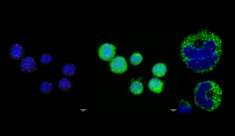
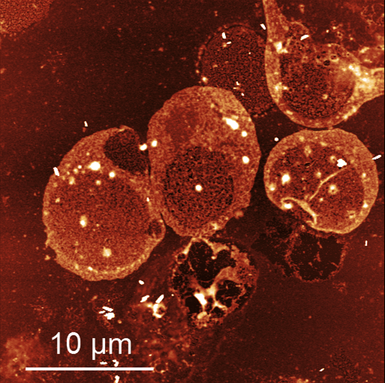
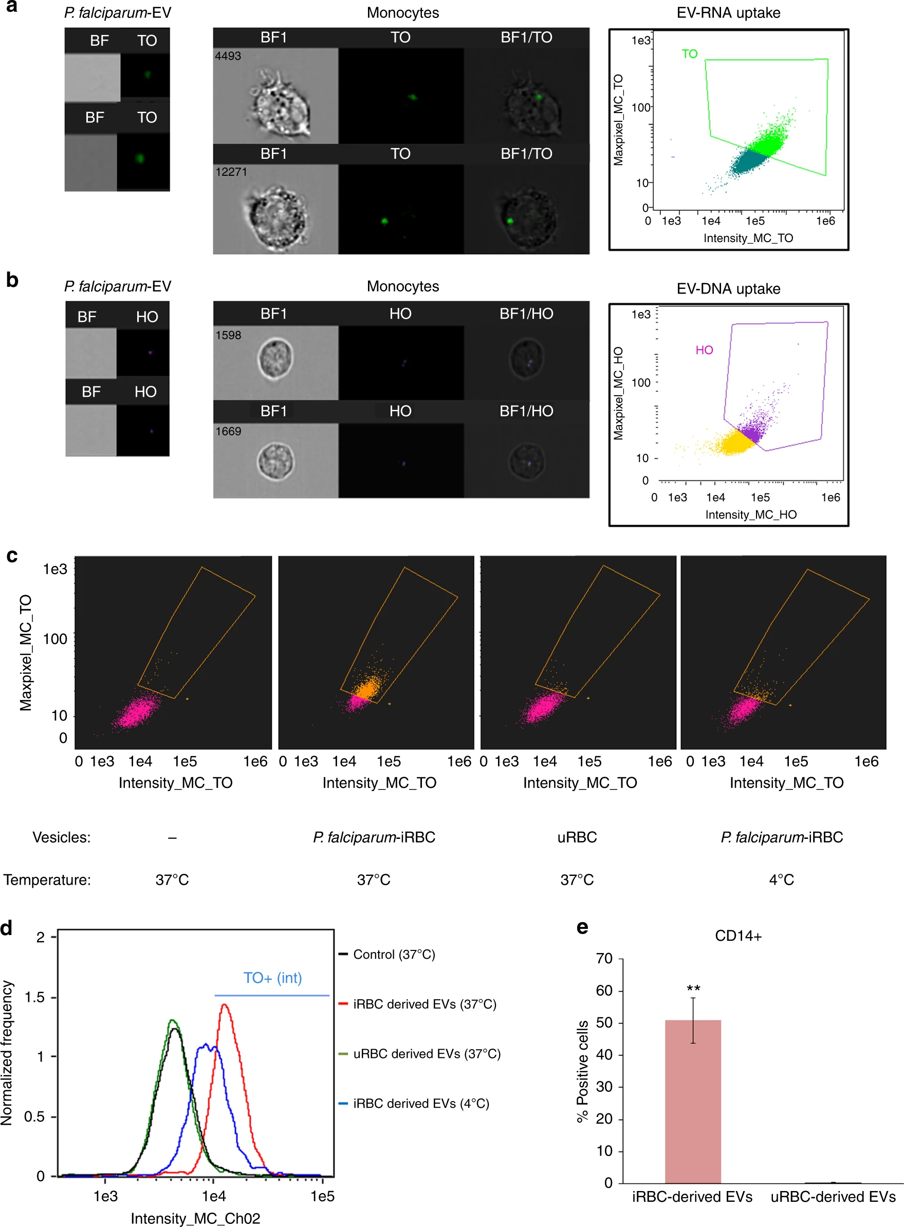
STING is an innate immune cytosolic adaptor for DNA sensors that engage malaria parasite (Plasmodium falciparum) or other pathogen DNA. As P. falciparum infects red blood cells and not leukocytes, how parasite DNA reaches such host cytosolic DNA sensors in immune cells is unclear. Here we show that malaria parasites inside red blood cells can engage host cytosolic innate immune cell receptors from a distance by secreting extracellular vesicles (EV) containing parasitic small RNA and genomic DNA. Upon internalization of DNA harboring EVs by human monocytes, P. falciparum DNA is released within the host cell cytosol, leading to STING-dependent DNA sensing. STING subsequently activates the kinase TBK1, which phosphorylates the transcription factor IRF3, causing IRF3 to translocate to the nucleus and induce STING-dependent gene expression. This DNA-sensing pathway may be an important decoy mechanism to promote P. falciparum virulence and thereby may affect future strategies to treat malaria.
-
(2017) PLoS Pathogens. 13, 8, e1006524. Abstract
Metabolic changes within the cell and its niche affect cell fate and are involved in many diseases and disorders including cancer and viral infections. Kaposi's sarcoma-associated herpesvirus (KSHV) is the etiological agent of Kaposi's sarcoma (KS). KSHV latently infected cells express only a subset of viral genes, mainly located within the latency-associated region, among them 12 microRNAs. Notably, these miRNAs are responsible for inducing the Warburg effect in infected cells. Here we identify a novel mechanism enabling KSHV to manipulate the metabolic nature of the tumour microenvironment. We demonstrate that KSHV infected cells specifically transfer the virus-encoded microRNAs to surrounding cells via exosomes. This flow of genetic information results in a metabolic shift toward aerobic glycolysis in the surrounding non-infected cells. Importantly, this exosome-mediated metabolic reprogramming of neighbouring cells supports the growth of infected cells, thereby contributing to viral fitness. Finally, our data show that this miRNA transfer-based regulation of cell metabolism is a general mechanism used by other herpesviruses, such as EBV, as well as for the transfer of non-viral onco-miRs. This exosome-based crosstalk provides viruses with a mechanism for non-infectious transfer of genetic material without production of new viral particles, which might expose them to the immune system. We suggest that viruses and cancer cells use this mechanism to shape a specific metabolic niche that will contribute to their fitness.
-
(2017) Seminars in Cell & Developmental Biology. 67, p. 83-90 Abstract
Infectious diseases are the leading cause of death of children worldwide, causing a tenacious and major public-health burden. The dynamic interplay between pathogens and their host is one of the most complicated themes of the disease progression. Pathogens excel in developing different means to facilitate cell-cell communication via secreted vesicles, among others. The released vesicles are involved in the transfer of biologically active molecules that induce phenotypic changes in the recipient cells. The messages within the vesicles are delivered to coordinate diverse processes, including virulence factor expression, differentiation state and control of their population density. Importantly, production of such vesicles promotes pathogen survival, as it provides a secure means of pathogen-pathogen communication and an ability to manipulate host responses for their own benefits. This review highlights intriguing findings, which show the important role of EVs in the social activity of pathogens, within and in between their communities. We further present examples of how pathogens use EVs to alter host immune and non-immune responses. Advancing our understanding of cell-cell communication in infectious diseases will be particularly useful to decipher the complexity of the crosstalk between pathogens themselves and their hosts, leading to the development of therapeutic strategies for fighting infectious agents. (C) 2017 Elsevier Ltd. All rights reserved.
-
(2017) PLoS Biology. 15, 6, e2002711. Abstract
Necroptosis is a regulated, nonapoptotic form of cell death initiated by receptor-interacting protein kinase-3 (RIPK3) and mixed lineage kinase domain-like (MLKL) proteins. It is considered to be a form of regulated necrosis, and, by lacking the "find me" and "eat me" signals that are a feature of apoptosis, necroptosis is considered to be inflammatory. One such "eat me" signal observed during apoptosis is the exposure of phosphatidylserine (PS) on the outer plasma membrane. Here, we demonstrate that necroptotic cells also expose PS after phosphorylated mixed lineage kinase-like (pMLKL) translocation to the membrane. Necroptotic cells that expose PS release extracellular vesicles containing proteins and pMLKL to their surroundings. Furthermore, inhibition of pMLKL after PS exposure can reverse the process of necroptosis and restore cell viability. Finally, externalization of PS by necroptotic cells drives recognition and phagocytosis, and this may limit the inflammatory response to this nonapoptotic form of cell death. The exposure of PS to the outer membrane and to extracellular vesicles is therefore a feature of necroptotic cell death and may serve to provide an immunologically-silent window by generating specific "find me" and "eat me" signals.
-
(2017) The Journal of infectious diseases. 215, 3, p. 378-386 Abstract
Schistosomiasis traditionally has been diagnosed by detecting eggs in stool or urine. However, the sensitivity of these examinations is limited, especially in travelers with a low worm burden. Serologic tests have a greater sensitivity, but their results remain positive regardless of treatment and thus cannot be used for follow-up of patients. We hypothesized that detection of worm microRNAs (miRNAs) in serum can overcome the drawbacks of the existing diagnostic methods.
-
(2017) Methods. 112, p. 157-166 Abstract
Malaria is the most devastating parasitic disease of humans, caused by the unicellular protozoa of the Plasmodium genus, such as Plasmodium falciparum (Pf) and is responsible for up to a million deaths each year. Pf life cycle is complex, with transmission of the parasite between humans via mosquitos involving a remarkable series of morphological transformations. In the bloodstream, the parasites undergo asexual multiplications inside the red blood cell (RBC), where they mature through the ring (R), trophozoite (T) and schizont (S) stages, and sexual development, resulting in gametocytes (G). All symptoms of malaria pathology are caused by the asexual blood stage parasites. Flow cytometry methods were previously used to detect malaria infected (i) RBCs, in live or fixed cells, using DNA (Hoechst) and RNA (Thiazole Orange) stains. Here, by using imaging flow cytometry, we developed improved methods of identifying and quantifying each of the four parasite blood stages (R, T, S and G). This technique allows multi-channel, high resolution imaging of individual parasites, as well as detailed morphological quantification of Pf-iRBCs cultures. Moreover, by measuring iRBC morphological properties, we can eliminate corrupted and extra cellular (dying) parasites from the analysis, providing accurate quantification and robust measurement of the parasitemia profile. This new method is a valuable tool in malaria molecular biology research and drug screen assays. (C) 2016 Elsevier Inc. All rights reserved.
-
(2017) Trends in Parasitology. 33, 1, p. 2-4 Abstract
During its life cycle, the malaria parasite must cope with a set of diverse environments and institute strategies to alter its host's responses. A recent study remarkably demonstrates how these parasites exploit red blood cell products, loading them into 'armed' secreted vesicles sent to manipulate their host's 'endothelium battlefront', thereby promoting malaria infection.
2014
-
(2014) Sub-Cellular Biochemistry. 78, p. 201-17 25487023. Abstract
Mitochondrial chaperones mediate and affect critical organellar processes, essential for cellular function. These chaperone systems have both prokaryotic and eukaryotic features. While some of the mitochondrial co-chaperones have clear homologues in prokaryotes, some are unique to eukaryotes and have no homologues in the chaperone machinery of other cellular compartments. The mitochondrial co-chaperones are required for protein import into the organelle and in enforcing the structure of the main chaperones. In addition to novel types of interaction with their senior partners, unexpected and essential interactions between the co-chaperones themselves have recently been described.
2013
-
(2013) Cell. 153, 5, p. 1120-1133 Abstract[All authors]
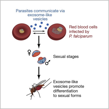
Cell-cell communication is an important mechanism for information exchange promoting cell survival for the control of features such as population density and differentiation. We determined that Plasmodium falciparum-infected red blood cells directly communicate between parasites within a population using exosome-like vesicles that are capable of delivering genes. Importantly, communication via exosome-like vesicles promotes differentiation to sexual forms at a rate that suggests that signaling is involved. Furthermore, we have identified a P. falciparum protein, PfPTP2, that plays a key role in efficient communication. This study reveals a previously unidentified pathway of P. falciparum biology critical for survival in the host and transmission to mosquitoes. This identifies a pathway for the development of agents to block parasite transmission from the human host to the mosquito.