Research

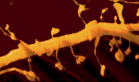
Calcium regulation of structural plasticity in cultured neurons

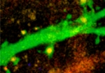
Hippocampal neuron taken from newborn rat, transfected with Orai1 (red) and GFP (green), grown for 2 weeks in culture, imaged at high resolution in a confocal microscope, to detect changes in dendritic spines (small appendages, about 1um in diameter, stained with Orai1 (yellow=green+red). (For further details, see 330)  

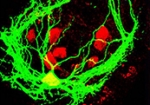
Small networks in island can generate synchronized activity

A small network grown on an island, containing eight cells (red). Activity of one of these neurons was recorded with patch clamp electrophysiological methods and was stained with biocytin (green) to allow detection of all the processes that the neuron sends out. These processes grow only within the boundary of the island, and the method allows detection of activity of all the neurons participating in the activity of the network. This allows analysis of the minimal network properties that can produce synchronous activity in controlled conditions. (For further details, see 290)

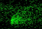
Fast confocal imaging of changes of calcium in dendritic spines

Line scan of changes in intracellular calcium in an individual dendritic spine following flash photolysis of caged calcium near dendritic spine head (red dot on the left). A line is scanned at a rate of ca. 1msec per line, from left to right, between spine head and parent dendrite, and intracellular calcium is raised momentarily (bottom left side), showing a rise in calcium (green fluorescence). This rise is spread from the point source of origin, throughout the dendritic spine, and into the parent dendrite. (For further details, see 291)