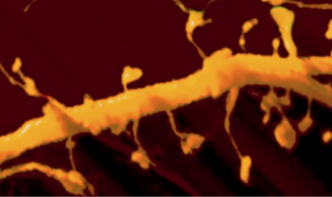
Line scan of changes in intracellular calcium in an individual dendritic spine following flash photolysis of caged calcium near dendritic spine head (red dot on the left). A line is scanned at a rate of ca. 1msec per line, from left to right, between spine head and parent dendrite, and intracellular calcium is raised momentarily (bottom left side), showing a rise in calcium (green fluorescence). This rise is spread from the point source of origin, throughout the dendritic spine, and into the parent dendrite. (For further details, see 291)

Head: Prof. Menahem Segal

