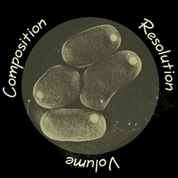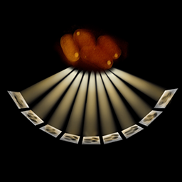

The living cell inspires us as a laboratory of nanoscale materials and molecular machines. Imaging and analytical tools such as optical tweezers were invented in order to investigate them. In recent years the lab is dedicated to development and application of scanning transmission electron microscopy (STEM) for cryo-tomography of cells and intra-cellular structures. The flexibility of STEM configuration allows optimization for diverse aims, including expanding the volume accessible for imaging in 3D and compositional contrast with elemental spectroscopy. Application examples include inorganic deposits in mitochondria, heme crystallization in Plasmodium, supramolecular protein assemblies, and large scale assemblies in chromatin.
Earlier works in the lab focused on molecular dynamics in the cell using primarily visible light imaging methods. Topics included protein exchange across the nuclear envelope (nuclear transport), structural biology of a gene transfer protein from Agrobacterium tumefaciens, synthetic protein self-assembly based on ferritin, and the crystallization of heme by the malaria-causing parasite Plasmodium falciparum.