

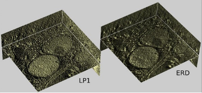
3D Deconvolution processing for tomography
In fluorescence microscopy, the haze due to out-of-focus light can be suppressed by deconvolution with a point spread function (PSF) that describes the imaging process. We show that STEM tomography can be described via a synthetic PSF constructed from the illumination profile tilted to the projection angles used in generating the reconstruction. Deconvolution is very effective in denoising the tomogram and in suppressing artefacts coming from the missing wedge of unacquired data.
Waugh et al. (2020) 3D Deconvolution Processing for STEM Cryo-Tomography. PNAS. http://www.pnas.org/content/117/44/27374
Recently the method was extended to TEM tomography.
Croxford et al. (2021) Entropy Regularized Deconvolution of Cellular Cryo-Transmission Electron Tomograms. https://www.biorxiv.org/content/10.1101/2021.04.26.441469v1
Advances in STEM for phase and depth contrast
Most classical STEM methods rely on incoherent contrast. In cryo-TEM of biological macromolecules, high resolution depends on coherent phase contrast. This motivates an exploration of different phase contrast (DPC) using a segmented diode detector (Opal, El-Mul Technologies Ltd, Israel). Using a custom-built scan generator and digitization system, we found that simultaneously-acquired STEM signals can be combined to produce images representing both phase and depth. Application to cryo-imaging is underway.
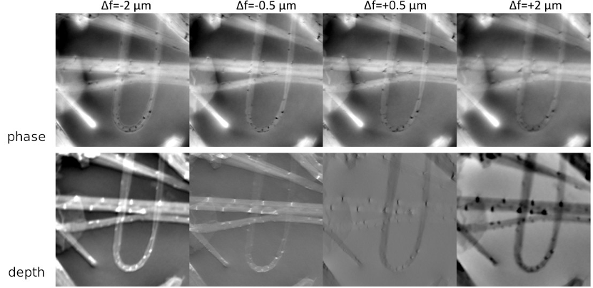
Seifer S., Houben L. & Elbaum M. (2021) Flexible STEM with Simultaneous Phase and Depth Contrast. Microscopy and Microanalysis. https://www.cambridge.org/core/journals/microscopy-and-microanalysis/article/flexible-stem-with-simultaneous-phase-and-depth-contrast/131F0EAA69FD8D9665E7E685EC81B1BE#