Covers
"The Brain": Cold Spring Harbor Symposia (1990)
Figure courtesy of Ts'o, Frostig, Lieke and Grinvald
See details
High resolution imaging of ocular dominance column based on intrinsic signals.
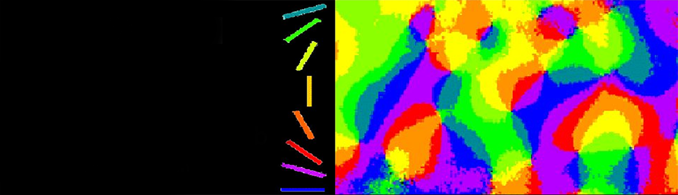
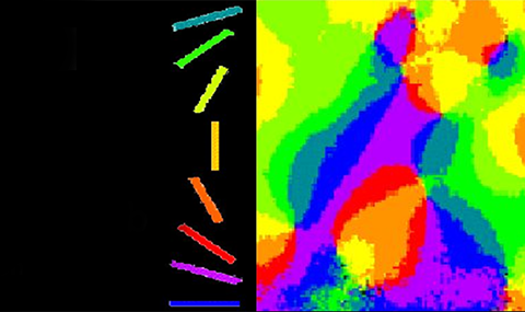
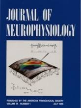
Organization of orientation and ocular dominance columns in the primate primary visual cortex
Daniel Y. Ts'o, Ron D. Frostig, Edmund E. Lieke, Amiram Grinvald.
Science Jul 27 1990, 249: 417-420.
Selective visualization of coherent activity in neuronal assemblies
Amos Arieli, Doron Shoham, Rina Hildesheim & Amiram Grinvald.
Journal of Neurophysiology, May 1995, 73: 2072-2093.
See details
For decades neurophysiologists have aspired to accomplish visualization of neuronal assemblies in action. A neuronal assembly may be defined as a group of neurons that cooperate to perform a specific computation required for a specific task. The activity of cells in an assembly is timelocked (coherent). However, the cells that comprise an assembly may be spatially intermixed with cells in other neuronal assemblies that are performing different computational tasks. The visualization of the spatio-temporal pattern of ongoing or evoked coherent-activity in neuronal assemblies has been accomplished by making use of the fact that activity of the neurons in an assembly is timelocked. The firing of a single neuron served as a time reference to selectively visualize only activity that was synchronized with it.
To study the spatio-temporal organization of neuronal assemblies single unit recordings and optical imaging based on voltage sensitive dyes were combined. Subsequently, spike-triggered averaging of the optical images relative to the spike of the firing neurons were performed. With sufficient averaging the activity of other neuronal assemblies not timelocked to the reference neuron was averaged out, thus enabling the selective visualization of the reference neuron's coherent assembly.
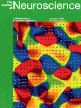
Functional organization for direction of motion and its relationship to orientation maps in cat area 18
Amir Shmuel and Amiram Grinvald.
Journal of Neuroscience, Nov 1 1996, 16: 6945-6964.
See details
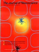
Map of the organization for direction of motion, featured on the November 1st, 1996 cover of the Journal of Neuroscience, by Amir Shmuel and Amiram Grinvald from the Weizmann Institute of Science.
Functional map for direction of motion in cat area 18. The colors code the local preferred direction of motion, also illustrated by the superimposed arrows. The length of the arrows is proportional to the local magnitude of direction selectivity.

"Imaging Neurons": A Laboratory Manual
Figure courtesy of Amiram Grinvald and Tobias Bonhoffe,r using material from Mark Hubener, Doron Shoham, Eyal Bartfeld and others
Book Editors R. Yuste, F. Lanni and A. Konnerth, Cold Spring Harbor Laboratory Press
See details
Optical imaging based on intrinsic signals enables mapping of the geometric relationship between various modules underlying visual processing in primary visual cortices of monkeys (the cube) and cats (the ellipse). The suggestions that geometric relationships exists (as schematically depicted on the surface of the cube) were first put forward by Hubel & Wiesel and Hubel & Livingstone in their famous ice-cube model. They were later revealed by intrinsic optical imaging (as schematically depicted on the surface of the cube). Optical imaging based on intrinsic signals can be combined with anatomical methods such as biocytin labeling of single neurons elucidating the relationship between neural structure and function (bottom left square). It also permits chronic recordings of functional maps providing new insights into the postnatal development of the neocortex or recording from behaving monkeys explaining higher brain functions.
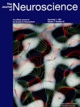
Spatial relationships among three columnar systems in cat area 17
Mark Hubener, Doron Shoham, Amiram Grinvald & Tobias Bonhoeffer.
Journal of Neuroscience, Dec. 1 1997, 17: 9270-9284,
See details
Map of the working brain obtained with optical imaging, featured on the December 1, 1997 cover of the Journal of Neuroscience.
- Black and white regions are columns of neurons that receive inputs from the left and right eye respectively. This system is responsible for depth perception.
- The 'pinwheel' spokes are formed by neuronal groups involved in the perception of shape, with each color line marking the border between neurons responsible for detecting a particular orientation in space.

Evolutions: Primary visual cortex
The Journal of NIH Research 5, (1993) 112
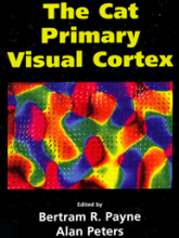
"The Cat Primary Visual Cortex"
Editors:Pertram R. Payne & Alan Peters.
Figure courtesy of Amir Shmuel & Amiram Grinvald
Spatiotemporal Dynamics of Sensory Responses in Layer 2/3 of Rat Barrel Cortex
Carl C. H. Petersen, Amiram Grinvald & Bert Sakmann.
Journal of Neuroscience, Feb 15, 2003, 23: 1298-1309
See details
Measured in vivo by voltage-sensitive dyeimaging combined with whole-cell voltage recodings and neuron reconstructions.

"Imaging in Neuroscience and Development"
Editors: Rafael Yuste and Arthur Konnerth.
Figure courtesy of Grinvald, Groner and Yuste (2004)
Neural Science - A century of progress and the mysteries that remain
Thomas D. Albright, Thomas M. Jessell, Eric R. Kandel, Michael I. Posner.
Figure courtesy of Amiram Grinvald, xxx
Neuron, Feb. 2000, Vol. 25, S1-S55.