Communication between the brain and the circulation
Communication between the brain and the circulation
In an attempt to understand how T cells can support the brain even though they are excluded from direct interaction with the brain parenchyma, we found that effector and regulatory CD4 T cells with specificity to CNS-antigens reside in the stroma of the brain’s choroid plexus [1, 2], and suggested the possibility that immune cells affect brain function from afar, via interactions with this compartment [3].
Choroid plexus dysfunction in brain aging
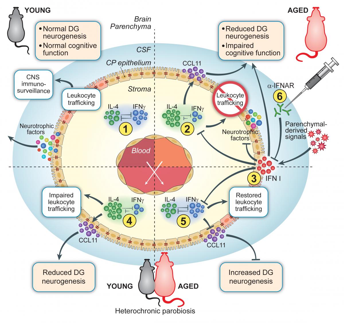
The CNS barrier system includes the blood–brain barrier (BBB), which is formed by tightly connected endothelium that surrounds parenchymal micro vessels, and the blood–CSF barrier (BCSFB), the barrier properties of which are established by the choroid plexus (CP), a highly vascularized epithelial monolayer that surrounds an inner stroma. While classically recognized for its role in the production of the cerebrospinal fluid (CSF), the CP is strategically positioned at the brain’s ventricles to be exposed to brain-derived signals from CSF, and to peripheral signals from the circulation. Examining the CP compartment throughout adulthood and aging in mice, we found that it undergoes immunological changes which affect its epithelial tissue [2]. Using multi-organ genome-wide analysis of young and aged mice, we further found that aging of the CP is characterized by a unique immunological signature of type I interferon (IFN-I) activity, which we also found in aged human brains [4]. Uncoupling the aging process of the immune system from that of the brain, by using heterochronic parabiosis model, in which aged mice are surgically connected to share vasculature with young mice, we showed that this response is induced by brain-derived signals which are present in the CSF, but not in the blood of aged mice. Additionally, we demonstrated that blocking IFN-I signaling within the aged brain can partially re-establish CP activity and, perhaps more importantly, ameliorate cognitive deficits and induce hippocampal neurogenesis in aged mice. These findings suggested immunomodulation of the CP as a novel approach in attenuating age-associated cognitive decline or other pathological conditions associated with chronic IFN-I within the CNS, often linked to cognitive dysfunction.
From a barrier to a selective gateway
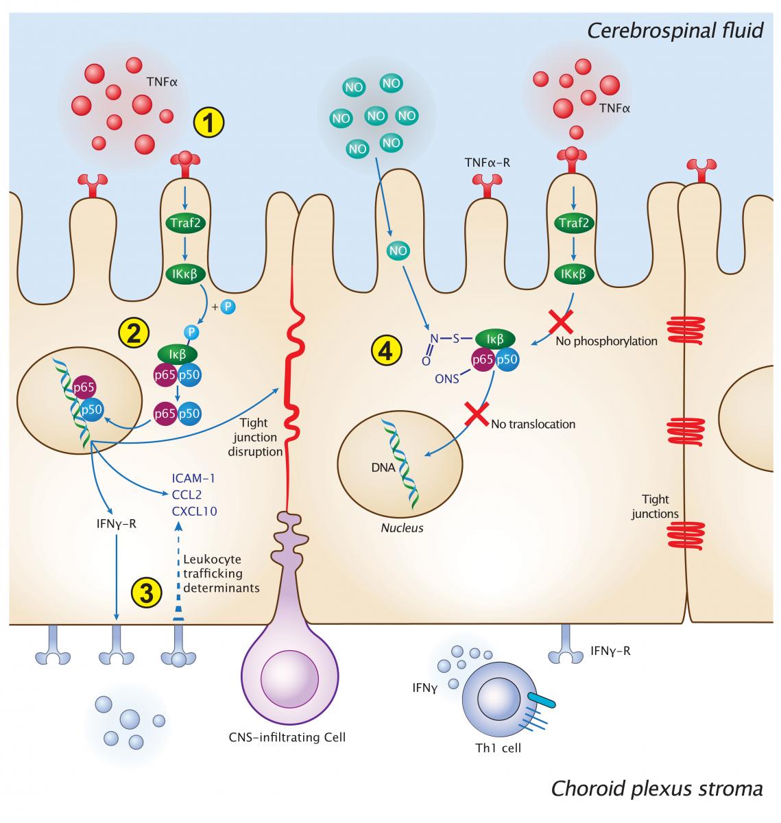
In contrast to the BBB, the BCSFB allows for immunosurveillance of the CSF under physiological conditions through the choroid plexus. Studying CP function in models of CNS acute injuries, we identified that it can function as a selective gateway for recruitment of monocyte-derived macrophages and T cells to the injured CNS parenchyma [1, 5]. Specifically, we found that CP activity in supporting immune cell trafficking dependents on a local synergistic effect in the CP compartment between CNS-derived inflammatory signals and the immune cell-derived cytokine interferon-γ [1], resulting in NFκB/p65-dependant CP gateway activity [6]. We also demonstrated how cerebroventricular elevation of nitric oxide elevation can negatively affect CP NFκB activation [6].
References
- Kunis G, Baruch K, Rosenzweig N, Kertser A, Miller O, Berkutzki T et al. IFN-gamma-dependent activation of the brain's choroid plexus for CNS immune surveillance and repair. Brain : a journal of neurology, 2013;136:3427-40.
- Baruch K, Ron-Harel N, Gal H, Deczkowska A, Shifrut E, Ndifon W et al. CNS-specific immunity at the choroid plexus shifts toward destructive Th2 inflammation in brain aging. Proceedings of the National Academy of Sciences of the United States of America, 2013;110:2264-9.
- Baruch K, Schwartz M. CNS-specific T cells shape brain function via the choroid plexus. Brain Behav Immun, 2013;34:11-6.
- Baruch K, Deczkowska A, David E, Castellano JM, Miller O, Kertser A et al. Aging. Aging-induced type I interferon response at the choroid plexus negatively affects brain function. Science, 2014;346:89-93.
- Shechter R, Miller O, Yovel G, Rosenzweig N, London A, Ruckh J et al. Recruitment of beneficial M2 macrophages to injured spinal cord is orchestrated by remote brain choroid plexus. Immunity, 2013;38:555-69.
- Baruch K, Kertser A, Porat Z, Schwartz M. Cerebral nitric oxide represses choroid plexus NFkappaB-dependent gateway activity for leukocyte trafficking. The EMBO journal, 2015;34:1816-28.