Research

New spin labeling approaches

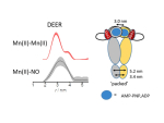
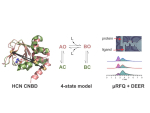
Electron paramagnetic resonance (EPR) techniques, specifically double electron-electron resonance DEER (or pulsed electron double resonance, PELDOR) are highly effective for determining the distances between two strategic sites in biomolecules such as proteins, nucleic acids, and their assemblies. These are usually carried out in frozen solutions and provide, for example, sparse structural information that can be used for tracking conformation changes upon ligand/substrate binding. At the heart of this methodology lies the controlled labeling of the molecules of interest with paramagnetic probes, between which the distances are measured. These could be either intrinsic paramagnetic centers such as transition metal ions and radicals or artificially introduced spin labels.

Observing protein’s conformational change in the cell

In-cell structural stability and the conformation of biomolecules might differ from in vitro conditions, owing to the complex, strong, or weak interactions they experience with other cell components as well as cellular crowding and post-translation modifications. Therefore, one has to ‘look’ at protein structure and dynamics inside the cell.

Conformational dynamics of Hsp90

Hsp90 is an essential molecular chaperone of the cell that modulates the folding, activation, and stability depending on ATP hydrolysis of a plethora of client proteins represented in many types of cancer. We are performing DEER distance measurements using non-conventional labeling schemes, including Gd(III), Mn(II), and nitroxides and their combinations, along with ENDOR experiments, to track the conformational changes of Hsp90 during the hydrolysis process in the presence of various co-chaperons and inhibitors.

Liquid-Liquid phase separation in proteins

Many membrane-less compartments in the cell, which behave as liquid-like droplets, also referred as coacervates, are formed via liquid-liquid phase separation (LLPS). Knowledge about the structural and dynamic properties of proteins that form membrane-less compartments is essential to understand the mechanism leading to LLPS and its associated intra- and inter-molecular interactions. 

Proteins at work – rapid freeze quench

Our microfluidic rapid freeze-quench design allows us to trap and investigate changes of protein conformation and structure in a time-resolved manner. The improves design is optimized for very small sample volumes, allowing the investigation of proteins that are difficult to obtain.

EPR methodology

In our group, we employ two home-built spectrometers, which are subject to continuous improvement. Additionaly, our research is focused towards the development and improvement of novel experiments, leading to increased sensitivity and applicability.