Research

Brain as a dynamic poro-viscoelastic biophysical system

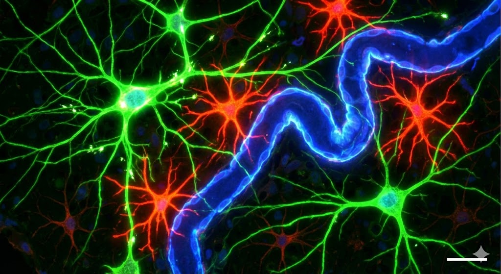
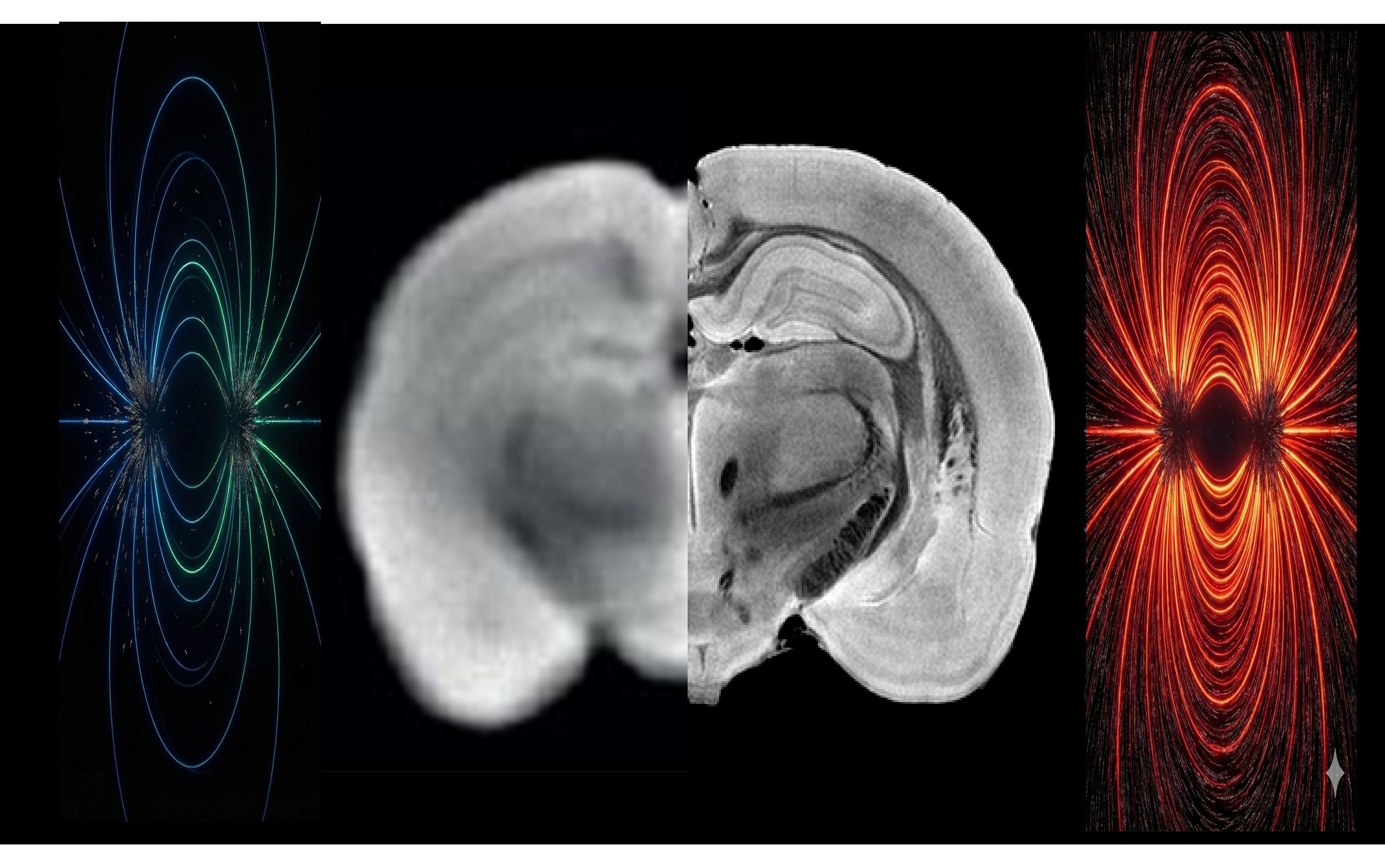
The brain is not just a signaling network — it is an active, deformable porous material whose microstructure and mechanics continuously interact with neural activity. We use living brain slices inside the MRI to link activity readouts (calcium imaging / electrophysiology) with dynamic microstructural and mechanical mapping using advanced MRI contrasts (diffusion, relaxation, elastography…) and advanced biophysical modelling. Using optogenetics, focused ultrasound, and targeted chemical perturbations, we causally manipulate both neural dynamics and tissue state to uncover feedback loops: how activity writes biophysical states, and how those states shape subsequent activity.

Read more

Mapping brain function with MRI beyond BOLD

BOLD-fMRI has transformed neuroscience, but it does not directly map neural activity, rather relying on vascular and metabolic coupling to deliver its signals. Thus, BOLD-fMRI can suffer from limiting specificity and interpretability, especially in disease where neurovascular coupling may be perturbed. We develop new functional MRI approaches and pulse sequences that probe neural activity and input/output properties more directly and via novel coupling mechanisms, including diffusion-based functional contrasts (dfMRI) and ionic/chemical contrasts such as sodium fMRI. Our methods are applied for mapping function in rodents (including in disease models) and validated in living brain slices, where vascular/BOLD effects are absent, enabling clean mechanistic grounding and direct validation against simultaneous optical/electrical activity measurements.

Read more

MR microscopy: pushing resolution in space and time

We develop ultrahigh field MR microscopy methodologies that push the limits of spatial resolution, temporal resolution, and quantitative inference. On the temporal side, we develop ultrafast fMRI acquisitions with advanced computational analyses and multimodal readouts to link understand information flow and oscillatory signals and their underlying neuronal drivers. On the spatial side, we develop ultrahigh-resolution in vivo MRI and analysis pipelines, aiming to resolve small cell clusters with multiple contrasts, and applying them to rodent models of plasticity and disease to track how evolves over time and how it relates to function. We further validate our work with advanced microscopy and histological approaches.

Read more