Methods

Targeted Patch-Clamp Recordings
We perform in vitro patch-clamp recordings from genetically identified retinal neurons. This approach allows us to measure synaptic inputs, intrinsic properties, and light-evoked responses with single-cell resolution.
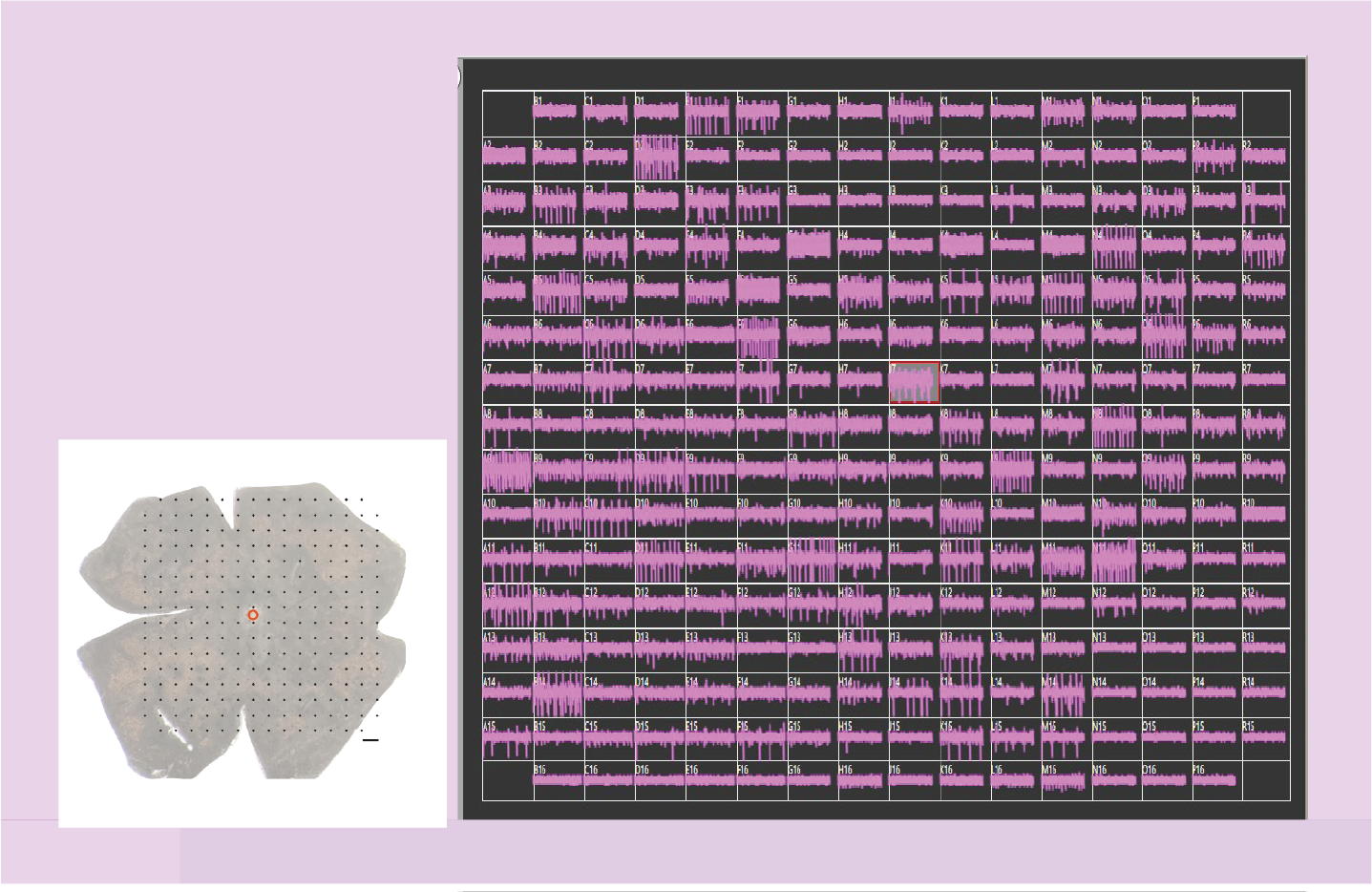
Multielectrode Array (MEA) Recordings
We use high-density MEA platforms to record the activity of hundreds of retinal ganglion cells simultaneously. This technique enables functional classification and population-level analysis of retinal output under diverse visual conditions.

Two-Photon Calcium Imaging
To monitor neural activity with high-spatial cellular resolution, we use two-photon Ca²⁺ imaging in transgenic or virally labeled tissue. This allows us to visualize population dynamics in the retina during visual stimulation.
Neuropixels Recordings
We use Neuropixels probes for large-scale, high-resolution electrophysiological recordings in vivo. These recordings track how visual information from the retina is represented and processed in downstream brain regions.
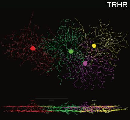
Histology and Immunostaining
We apply immunohistochemistry and confocal imaging to visualize retinal cell types and dendritic morphology supporting circuit-level analysis and validation.
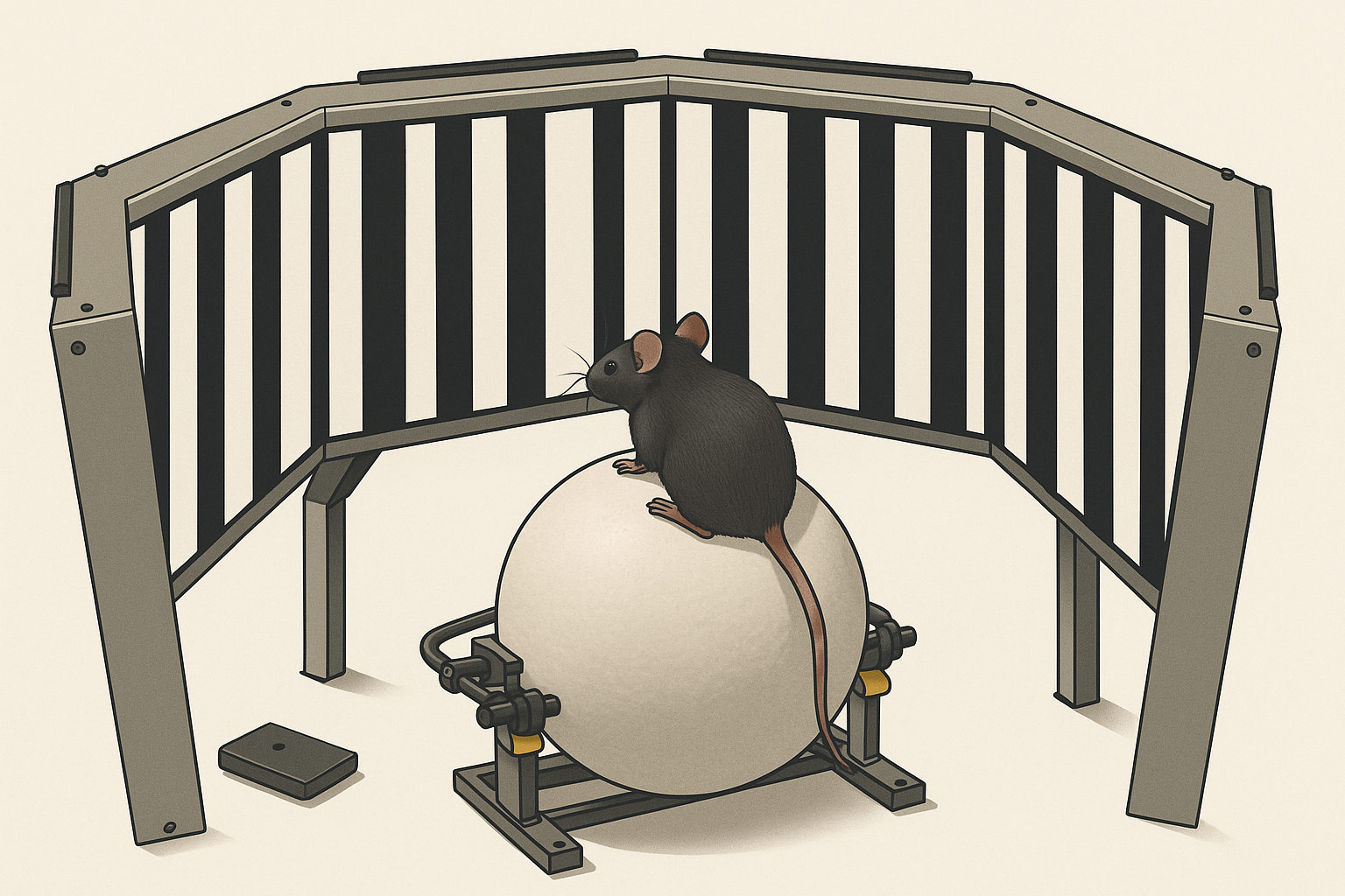
Behavioral Assays
We design behavioral tasks to test how retinal computations influence perception, learning and action. Combining behavior with genetic and electrophysiological tools helps link retinal function to natural and learned visual behaviors.

Viral Tools and Genetic Manipulation
We use AAVs to label and manipulate neuronal circuits, enabling targeted monitoring and control of cell types for studying visual computation.