Topographic Variation in Retinal Computations
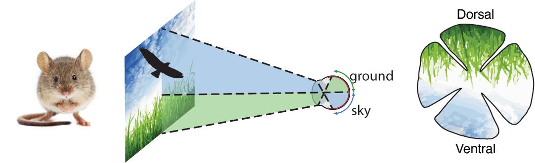
Retinal ganglion cells (RGCs) of the same type are typically assumed to respond similarly, regardless of their position in the retina. However, animals view the world through species- and location-specific perspectives. We've shown that RGCs of a single type can exhibit distinct response properties across retinal locations, suggesting that visual encoding is tuned to the behavioral demands of different parts of the visual field.
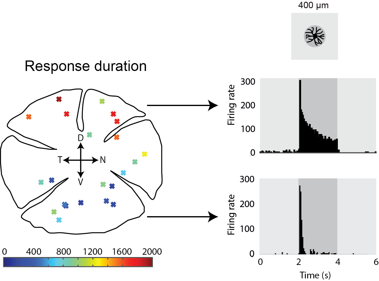
We found that transient-Off-α RGCs respond differently depending on their retinal location: in the ventral retina, responses to dark stimuli are brief and transient, while in the dorsal retina, they become over five times longer in duration. Using intracellular recordings, pharmacology, and genetic tools, we showed that this variation is driven by location-dependent differences in synaptic input from the primary rod pathway. These results suggest that retinal circuits are evolutionarily tuned to local visual environments, optimizing how different regions of the retina sample the world.
See more:
Warwick et al., 2018, Inhomogeneous Encoding of the Visual Field in the Mouse Retina
Heukamp, Warwick et al., 2020, Topographic Variations in Retinal Encoding of Visual Space