


Magnetic resonance imaging (MRI) is a highly versatile, non-invasive tool for three dimensional visualization of in-vivo organs and tissues. A special role in MRI is played by so-called “ultrafast” techniques like Echo-Planar Imaging, EPI, capable of delivering these data in a fraction of a second. Our group works on developing new approaches to collecting ultrafast MRI data, which will provide an increased robustness over existing alternatives. Our new methods derive on the spatio-temporal encoding (SPEN) principles that we have developed for collecting arbitrary multidimensional NMR/MRI data in a single scan. Besides the physical development of these SPEN variants (e.g., upper right) we employ these methods to tackle important problems such as diffusion measurements to diagnose cancer (center) and functional MRI (fMRI) to monitor brain activities.
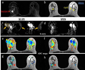
Breast Cancer Patient – Detection of malignancies by SPEN-based Diffusion Imaging (3 Tesla): comparison between images derived from spin-echo EPI and single-slice SPEN scans of a patient with IDC. Red arrowheads indicate the cancer; yellow arrowheads indicate the cysts; dashed orange regions highlight the folding of cysts and fat
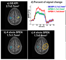
Functional SPEN MRI – Motor Activation Monitored in the Human Brain (3 Tesla) : fMRI based on motor stimuli including all fingers tapping for 30 seconds, interleaving right and left hands for a 5 minutes total study duration (5 pairs of stimuli blocks).
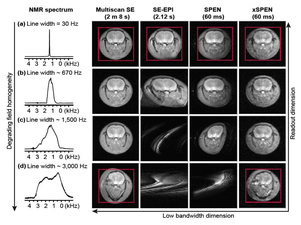
Performance of xSPEN 2D imaging is compared to other technique on an ex-vivo rat brain sample at 7 Tesla. Ex-vivo rat brain 1D NMR and 2D imaging results from different sequences, collected for increasingly degraded magnet field homogeneity conditions. (a) 1D spectrum and images acquired using multi-scan SE, SE-EPI, SPEN and xSPEN sequences, in a uniform magnetic field where the water resonance is characterized by a 30 Hz full width at half maximum. (b, c, d) Idem but acquired in magnetic fields that were deliberately and arbitrarily deteriorated, as evidenced by the increasing 1D NMR half-height line widths mentioned on the left-hand column. Notice the progressive distortions introduced by the field inhomogeneity in all images but the xSPEN one, as judged by the dotted squares added for ease of viewing.