
Reference-Based MRI
L. Weizman and Yonina C. Eldar
Introduction
MRI is the method of choice for clinical brain imaging, as it involves no exposure to ionizing radiation and provides high quality of soft tissue imaging. Conventional clinical MRI scan may last up to one hour and consists of multiple imaging contrasts of the same region. Radiologists are able to detect subtle abnormalities, such as a developing tumor, by comparing multiples images of the same region and examining the variations in contrast of the different tissue types. However, the scanning procedure in MRI is time-consuming, resulting in patients spending prolonged times inside the machine.
To substantially shorten the MRI scanning times and improve SNR we propose a general framework that utilizes a reference image that exists in many MRI imaging scenarios. This reference can take on different forms: different imaging contrasts as done routinely in MRI acquisition, adjacent slices, or prior scans of the same patient. In all of these cases we can use the prior information in an adaptive fashion.
As a result MRI scans are much faster completed and can lower the time that patients have to spend inside the machine from almost one hour to just 15 minutes!
Algorithmically, the reference is incorporated by using a general framework which relies on solving the following optimization problem:
where y is the acquired data is the k-space domain, x is the imaging data to reconstruct and x0 is the reference scan data (assumed to be similar to x. Ψ is a sparsifying transform operator and Fu is undersampled Fourier operator. A controls the weight given to the fidelity of certain measurements (used to prioritize samples with high SNR).
The matrices W1and W2 depend on the application and control the weight given to each element in the sparse representation. In particular, W2 is used to weight image regions according to their similarity level with the reference scan. The parameters λand λ2 are regularization parameters that control the weight given to each term in the optimization problem
Applications
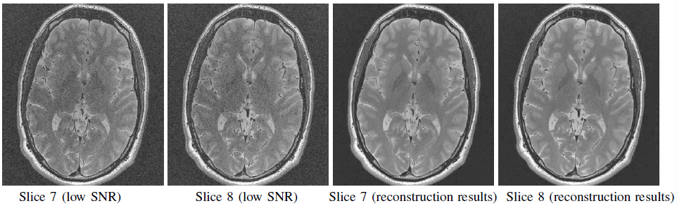
SNR improvement within the same imaging contrast: In MRI, SNR is proportional to the number of protons involved in generating the measured signal. As a result, thick slices provide better SNR than thin ones. To provide high SNR in 3D scans consisting of thin slices, scanning has to be repeated and averaged over several repetitions. In this application, adjacent MRI slices are used are reference images. We acquire low SNR scan consisting of thin slices, and high SNR scan consisting of thick slices where each thick slice overlaps two thin ones. Our goal is to reconstruct a high SNR scan comprised of thin slices from this data. For this purpose we rely on the similarity between adjacent thin slices. Taking into account the we used only single repetition for reconstruction vs. four repetitions required for high SNR thin slices, yielding a speedup factor of 2.6. The figure below shows representative results of this application.
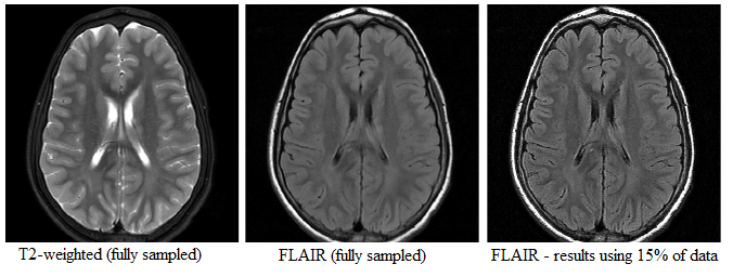
Utilizing similarity between imaging contrasts in same scan: MRI scan consists of multiple imaging contrasts. In many cases, we find similarity between two different imaging contrasts. In particular T2-weighted and fluid-attenuated inversion recovery (FLAIR) contrasts exhibit high similarity in non-fluid regions. In this application, the reference image is a different imaging contrast of the patient from the the same scan. Our goal is to reconstruct the FLAIR image, from undersampled measurements, given the T2 image as a reference scan. The figure below shows the similarity between T2 and FLAIR, and the reconstruction results of this application.
Utilizing similarity between repeated scans: Repeated brain MRI scans of the same patient every few weeks or months are very common for follow-up of brain tumors. The figure below shows the similarity between MRI scans of the same patient acquired at different dates.
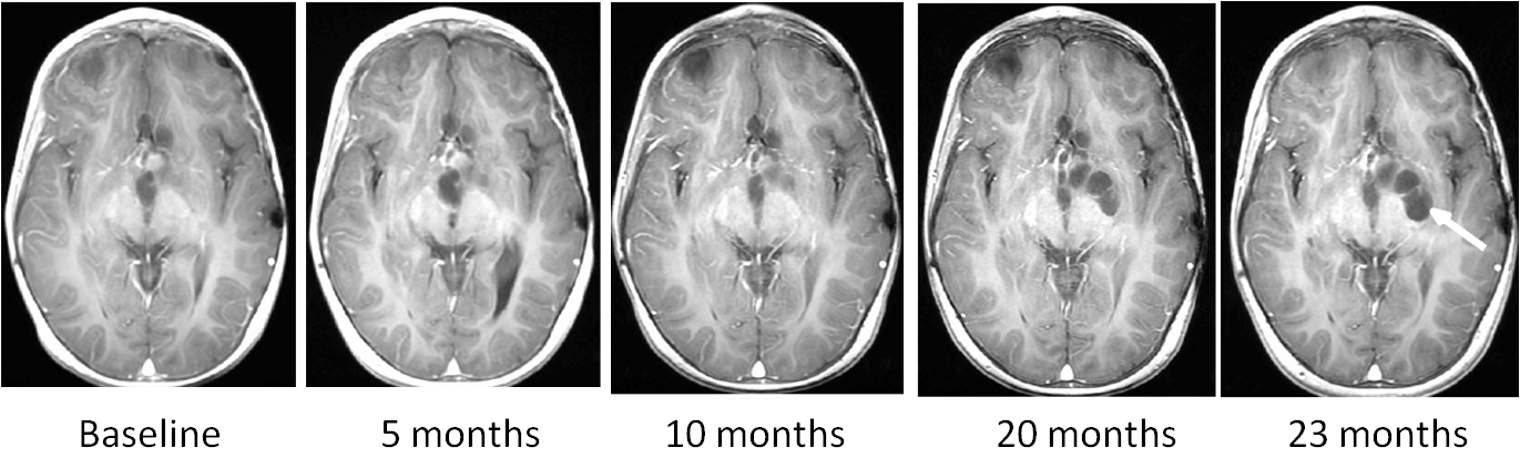
In this application, the reference scan is a previous scan of the patient in the time series, which is utilized for fast acquisition of a follow-up scan. In this application we need to address problems that do not exist in previous applications, since similarity between time points is not guaranteed. We tackle the unique properties of this application via an adaptive sampling mechanism.
References
L. Weizman, O. Rahamim, R. Dekel, Y. C. Eldar and D. Ben-Bashat, "Exploiting Similarity in Adjacent Slices for Compressed Sensing MRI", the 36th Annual International Conference of the IEEE Engineering in Medicine and Biology Society (EMBC), 2014.
L. Weizman, Y. C. Eldar and D. Ben Bashat, "Compressed sensing for Longitudinal MRI: An adaptive-weighted approach", Technical report, 2015.
Software Download
- Download the reference-based MRI SNR improvement package.
- Download the reference-based MRI Fast FLAIR package.
- Download the reference-based MRI Fast repeated scans package.
Installation:
1. Unzip each files to a separate empty directory.
2. Follow the instructions in readme.txt .