Mechanisms of axon pruning

Mechanisms of axon pruning

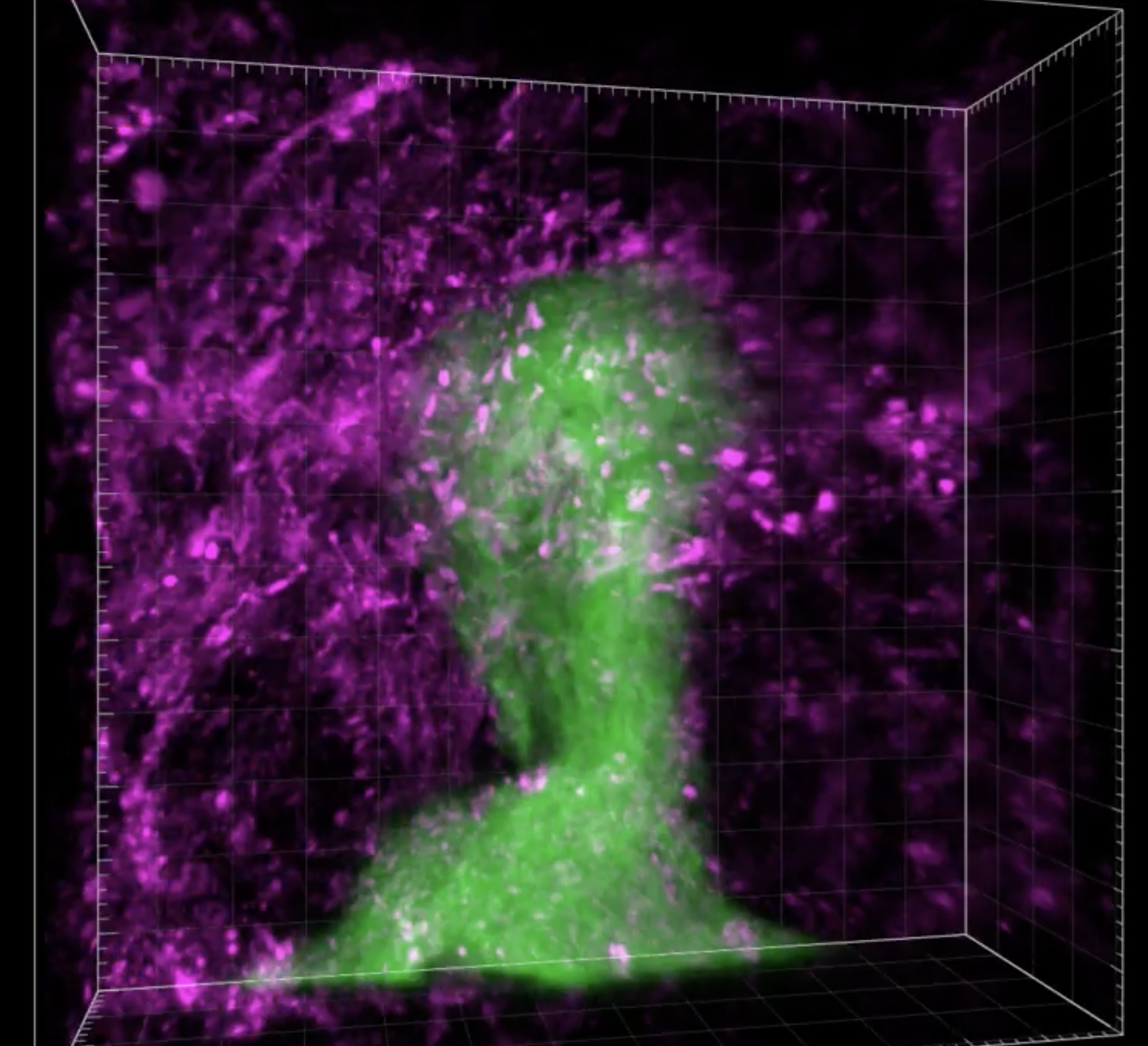
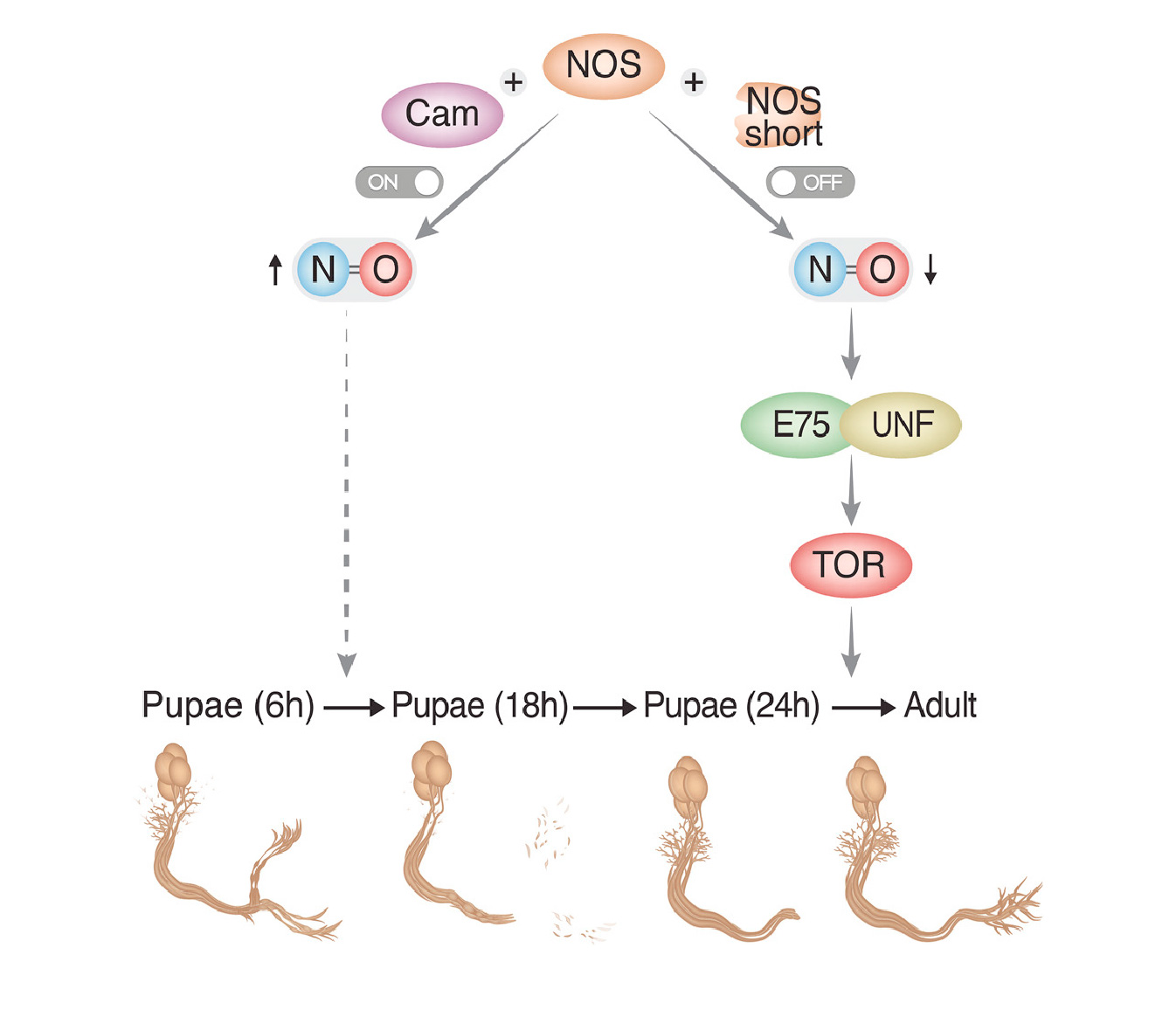
Developmental axon pruning of mushroom body (MB) γ neurons in the Drosophila fruit fly is a perfect model system to study neuronal remodeling: Not only can we harness the awesome genetic power of the fly and its short life cycle but we can also view the stereotypical neuronal remodeling as they occur in vivo during the larva to adult metamorphosis (see Figure 1). In our lab we use genetic tools  to explore the roles of intracellular signaling, trafficking and cytoskeletal changes during neuronal remodeling. In addition, we employ genomic tools to characterize in high resolution the transcriptional landscape of γ neurons at different stages of development.

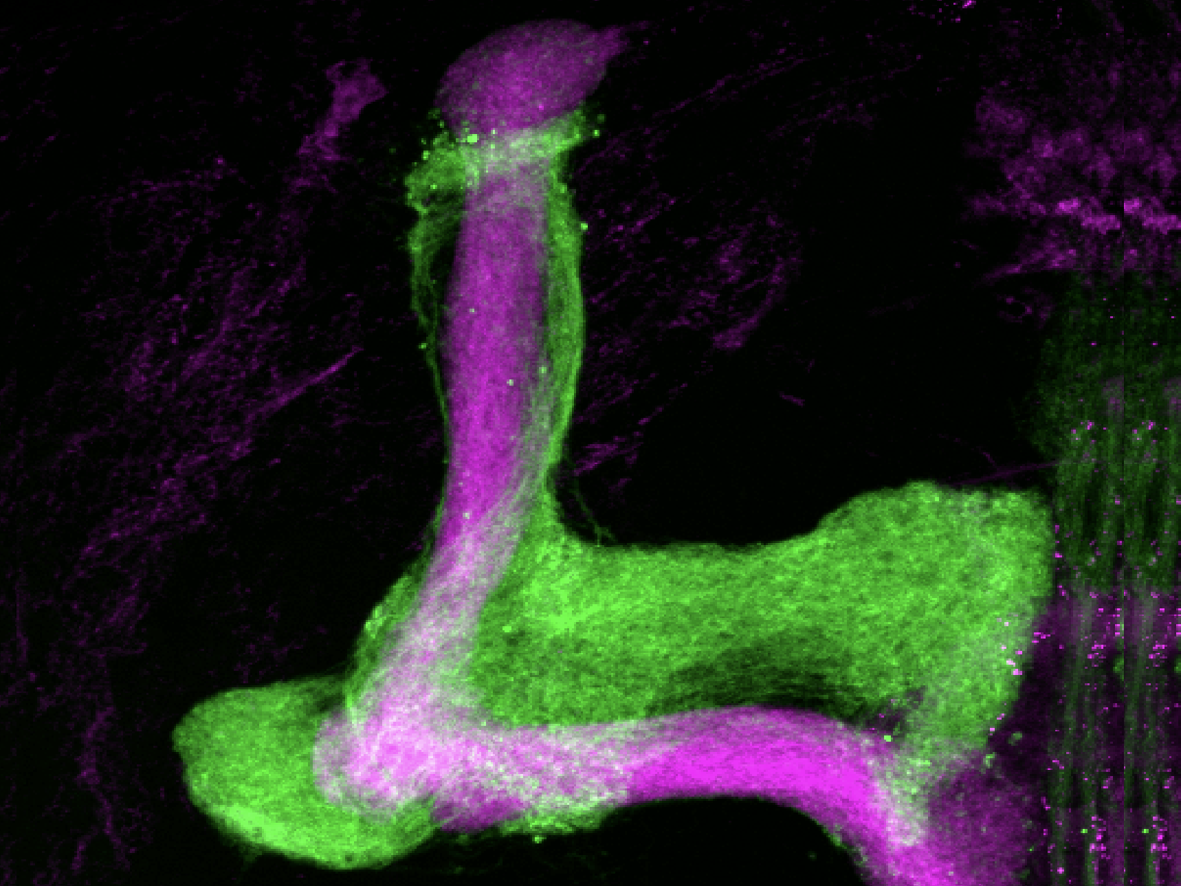
Figure 1: Neuronal remodeling of Drosophila Mushroom Body (MB) γ Neurons. (A) Schematic summary of MB development, indicating the three types of neurons generated from a common neuroblast precursor at three developmental stages. γ (red), α’/β’ (green) and α/β (blue) neurons have distinct axonal projection patterns in the adult brain. NHL, newly hatched larvae; ALH, after larval hatching; APF, after puparium formation.(B) Schematic representation and (C-D) γ neuron single/two-cell MARCM clones at different stages during development. (B,C) Larval neurons project axons to both the dorsal (d) and medial (m) lobes. (B,D) At 18 hr APF, axons prune larval-specific dorsal and medial branches (dashed arrows indicate the absence of the larval branches). (B,E) Adult neurons re-extended only medial (solid vertical arrow) but not dorsal (dashed horizontal arrow) branches. den, dendrites; p, peduncle; Green in (C-E) represents MB γ neuron MARCM clones labeled with mCD8-GFP; magenta represents anti-FasII staining, which labels strongly α/β neurons, weakly γ neurons, and does not label α’/β’ neurons. Modified from Watts et al (2003).