
Super Resolution in Ultrasound and Microscopy
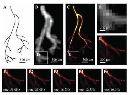
The attainable resolution of ultrasonography and optical imaging is fundamentally limited by diffraction, where the smallest resolvable feature is roughly half the wavelength. Improving resolution can greatly benefit applications such as imaging blood vessels (<100 μm) or cellular organelles (<100 nm).
In our lab, we use sparse frame sequences and model-based learning to surpass this limit. Inspired by a Nobel Prize-winning concept, we localize emitters at sub-diffraction precision and recover the underlying structure across frames. Unlike classical approaches that rely on frame-level separability, our learning-based methods can resolve dense, overlapping structures using fewer frames and in real time.
Our deep-unfolded networks integrate physical models and optimization principles for efficient recovery. These systems support single-frame learning, operate under challenging conditions, and generalize to diverse modalities—from live-cell microscopy to in-vivo ultrasound imaging. As a result, we enable practical super-resolution tools for both biological and clinical applications
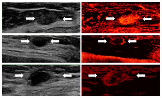
Super-resolution demonstrations in human ultrasound scans
show the vascular characteristics of three types of breast lesions:
- Top (Fibroadenoma - benign): The super-resolved image reveals an oval, well-circumscribed mass with uniform, dense vascularization.
- Middle (Cyst - benign): The recovery highlights peripheral vascular concentration surrounding a round, avascular core.
- Bottom (Invasive Ductal Carcinoma - malignant): An irregular, ill-defined mass with sparse central and dense peripheral vasculature.
Super-resolution vascular imaging of three breast lesions: fibroadenoma (top), cyst (middle), and invasive ductal carcinoma (bottom).
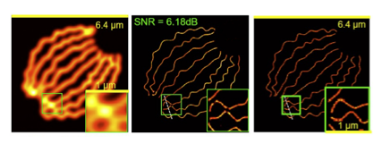
Super-resolution microscopy of biological tubulins:
Reconstruction of microtubule structures using 361 high-density frames. The left panel shows a diffraction-limited image; the middle panel presents our learning-based reconstruction, closely matching the right panel ground truth, demonstrating enhanced contrast and sub-diffraction detail recovery.
Tubulin structure recovered from high-density frames. Left: diffraction-limited image. Middle: our reconstruction. Right: ground truth.
References
- O. Bar-Shira, A. Grubstein, Y. Rapson, D. Suhami, E. Atar, K. Peri-Hanania, R. Rosen, Y. C. Eldar, "Learned Super Resolution Ultrasound for Improved Breast Lesion Characterization", 24th International Conference on Medical Image Computing and Computer Assisted Intervention (MICCAI 2021), October 2021.
- G. Dardikman-Yoffe and Y. C. Eldar, "Learned SPARCOM: Unfolded Deep Super-Resolution Microscopy", Optics Express, vol. 28, issue 19, pp. 27736-27763, September 2020.
- A. Bar-Zion, O. Solomon, C. Tremblay-Darveau, D. Adam and Y. C. Eldar, "SUSHI: Sparsity-Based Ultrasound Super-Resolution Hemodynamic Imaging", IEEE Transactions on Ultrasonics, Ferroelectrics and Frequency Control, vol. 65, issue 12, pp. 2365-2380, December 2018.
- O. Solomon, R. J. G. van Sloun, H. Wijkstra, M. Mischi and Y. C. Eldar, "Exploiting Flow Dynamics for Super-Resolution in Contrast-Enhanced Ultrasound", IEEE Transactions on Ultrasonics, Ferroelectrics, and Frequency Control, vol. 66, issue 10, pp. 1573-1586, October 2019.
-
R. J. G. van Sloun, O. Solomon, M. Bruce, Z. Z. Khaing, H. Wijkstra, Y. C. Eldar and M. Mischi, "Super-Resolution Ultrasound Localization Microscopy through Deep Learning", IEEE Transactions on Medical Imaging, vol. 40, issue 3, pp. 829-839, March 2021.