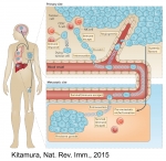
Research

Introduction

Circulating immune cells and hematopoietic progenitors must exit blood vessels near specific target sites of injury, inflammation or tissue repair. The vessel wall at these sites displays specific combinations of traffic signals in the form of adhesion molecules (selectins, integrins) and chemotactic cytokines (chemokines) which operate in sequence to recruit only specific circulating subsets with proper receptors to these signals. As these processes take place under shear stress, these traffic molecules have evolved to operate under specialized kinetic and mechanical contexts. 

The role of DC ICAM-1 in immune synapses in vivo

ICAM-1 is major cell adhesive ligand of the leukocyte integrin LFA-1. It is expressed on various vascular cells, pericytes, on different leukocytes including antigen presenting dendritic cells (DCs), macrophage subsets and B cells, as well as on various transformed epithelial cells. We have recently found that functional contacts between CD4 lymphocytes and adjuvant stimulated dendritic cells (DCs) inside lymph nodes (a model for skin vaccination) take place independently of ICAM expression on these DCs. Our work shows that priming and Th1 differentiation of these lymphocytes depend primarily on TCR signals rather than on T cell integrins. 

Targeting killer T cells to metastatic lesions

Although it is well established that tumor-specific CTLs enter primary tumors (e.g., raised against patient specific neoantigens or tumor-enriched antigens), the ability to use similar tumor-specific CTLs to eradicate secondary tumors (metastasis) at remote organs has been very limited because these injected CTLs must be able to exit (extravasate) blood vessels nearby the metastatic lesions they are assigned to eliminate. The lung is a major organ of metastasis of multiple malignancies including melanoma, breast cancer and lung cancer. Currently, the feasibility of targeting tumor specific killing T cells through the dense vasculature of the lung to enter metastatic lesions and kill these lesions is unclear.

The nuclear lamina of leukocytes and solid tumor cells: implications for migration, extravasation, proliferation and lung metastasis

Nuclear stiffness is controlled by the ratio between different lamins, major intermediate filament constituents of the nuclear lamina. Transendothelial migration of lymphocytes and neutrophils is associated with the ability of their deformable nuclei to displace endothelial cytoskeletal barriers. Lamin A is a key intermediate filament component of the nuclear lamina which regulates nuclear stiffness.

Lung infection and inflammation

The lungs are one of the most immunologically active organs in the human body. We are interested in dissecting the vascular signals that control leukocyte migration and function in clearing viral and bacterial infections, with a focus on influenza infection and vaccination. We have also established in vivo murine experimental models for smoke and LPS induced airway inflammation. We unraveled major protective roles of the tumor suppressor DAPk both in the context of LPS and smoke induced lung inflammation.

Breast cancer killing and metastasis

Malignant breast epithelial cells express elevated levels of the leukocyte integrin ligand ICAM-1. Tumor cells are targeted by NK cells and cytotoxic T cells (CTLs). These cells generate specialized killing synapses with both cancer cells and several stromal cells in their surroundings. Various studies implicated ICAM-1 on target tumor cells and LFA-1 or related integrins on killer leukocytes as essential for the generation of killing synapses but these killing synapses have been studied mainly in vitro focusing on malignant immune cells as targets rather than on solid tumors. Thus, evidence that this ligand is involved in the killing of specific solid tumors such as breast carcinoma by NK cells and CTL is still missing.  

Chemokine receptors and adaptors on microvilli

Our earlier work has shed light on the earliest signals transmitted by chemokines through their G-protein coupled receptors (GPCRs) to distinct leukocyte integrins (Scheme 1). In lymphocytes, the integrin adaptor talin1 was found to control major conformational changes of both LFA-1 and VLA-4 integrins implicated in lymphocyte arrest on endothelial ligands.

Chemokine vesicles

Chemokine vesicles

Endothelial-presented chemokines are critical for integrin-dependent adhesion and transendothelial migration (TEM) of naïve and memory lymphocytes. TH1 and Tc1 effector lymphocytes are special subsets of T cells that were recently activated by antigenic signals and underwent massive proliferation before they can exit the lymph nodes in which antigen was encountered. These cells were found by us to express adhesive integrins that can bypass chemokine signals and establish firm arrests on variably inflamed endothelial barriers.

Invasive filopodia

Invasive filopodia

Endothelial chemokines are instrumental for integrin mediated lymphocyte adhesion and TEM through vascular barriers. Addressing how chemokine signals promote these two processes using flow chamber assays, we found that following arrest, both T and B lymphocytes rapidly crawl on inflamed endothelium in an LFA-1 and chemokine dependent manner. We also found that endothelial presented chemokines trigger lymphocyte crawling by stimulating high affinity (HA) LFA-1 at numerous focal submicron dots underneath the entire adhesive zone reversibly generated by the rapidly crawling lymphocyte.

Mechanical barriers for leukocyte transmigration

Mechanical barriers for leukocyte transmigration

Leukocyte transendothelial migration (TEM) through inflamed endothelial cells (ECs) is initiated by leukocyte extension of a large leading edge under the endothelial barrier (basolateral lamellipodia) into which leukocytes squeeze their nuclei and thereby generate micron-sized gaps, primarily in between ECs (paracellular TEM). Our electron microscopy analysis suggests that ECs contain a dense cortical actin cytoskeleton which imposes a mechanical barrier for this dramatic squeezing process.

TCR activation

Lymphocyte stoppage on dendritic cells (DC) requires activation of LFA-1 by T cell receptor (TCR) signals, but the molecular basis of this activation is elusive. Using antibodies for specific LFA-1 conformations, we found that TCR activation in resting lymphocytes was insufficient to trigger LFA-1 extension or headpiece opening. However, TCR signals facilitate rapid conformational activation of LFA-1 by immobile ICAM-1, and promote lymphocyte spreading without requirement for cytosolic Ca2+.