
Contactless Vital Signs Radar

Outline:
-
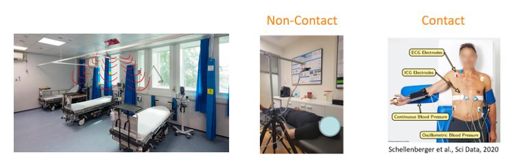
Non-contact vital signs monitoring (NCVSM) of multiple people in noisy, cluttered environments
-
Extension to NCVSM during large movements
-
RaLU-Net: Deep unfolded radar localization of humans
-
Non Contact Spirometry Test
-
Non-Invasive Quantitative Monitoring of Nanoparticles In Vivo
Non-contact vital signs monitoring (NCVSM) of multiple people in noisy, cluttered environments
Background:
Non-contact vital signs monitoring (NCVSM) of multiple individuals, such as respiration and heartbeat, has been investigated in recent years due to the rising cardiopulmonary morbidity, the risk of disease transmission, patient discomfort and heavy burden on medical staff. Frequency Modulated Continuous Wave (FMCW) radars have shown great promise in meeting these needs. However, contemporary techniques for NCVSM via FMCW radars, are based on simplistic models and present difficulties in coping with noisy environments containing multiple objects.
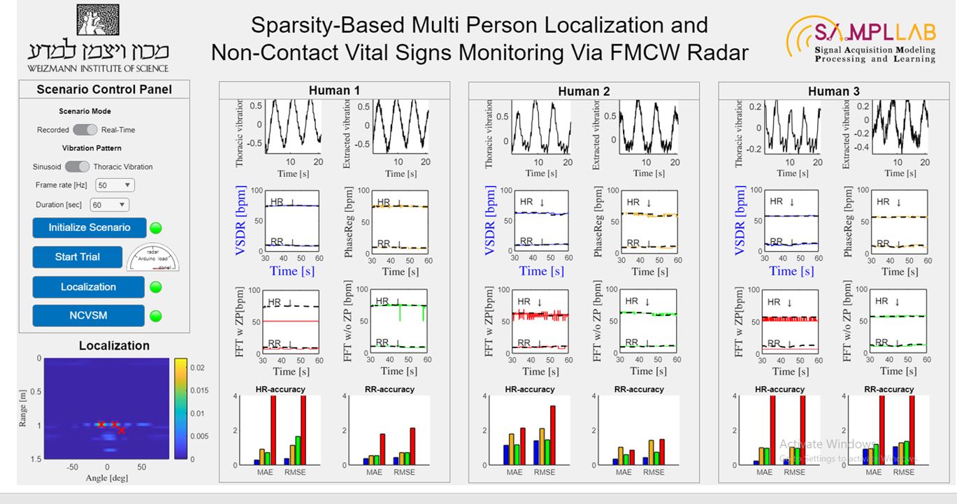
Sparsity-Based Multi-Person Localization and NCVSM
We developed an extended model for multi-person NCVSM via FMCW radar. By utilizing the sparse nature of the modelled signals in conjunction with human-typical cardiopulmonary features, we present accurate localization and NCVSM of multiple individuals in a cluttered scenario, using only a single channel. Specifically, we provide a joint-sparse recovery (JSR) mechanism to localize humans in a noisy environment and develop a robust method for NCVSM called VSDR (for vital signs based dictionary recovery), which uses a dictionary-based approach to search for the rates of respiration (RR) and heartbeat (HR) over high-resolution grids corresponding to human cardiopulmonary activity.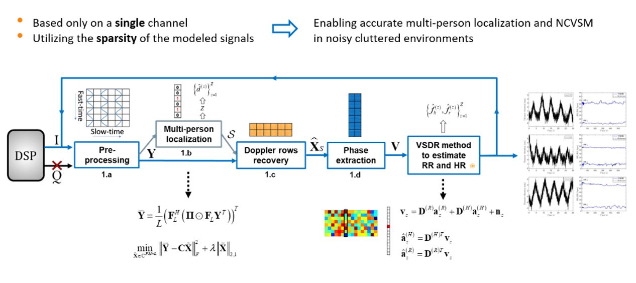
Fig. 1. Block diagram of the proposed approach for multi-person NCVSM via SISO FMCW radar, which yields accurate estimates of RR and HR by exploiting the sparse composition of the input data in accordance with human cardiopulmonary properties, using only a single channel.
The proposed approach was developed for SISO, SIMO and MIMO configurations.
SISO – single-input-single-output
SIMO – single-input-multiple-output
MIMO – multiple-input-multiple-output
Results
The advantages of our method are illustrated through comprehensive experiments as well as simulated data.
Experimental analysis:

Simulated analysis:
The simulated trials combine the proposed model with in-vivo data of 30 monitored individuals. We demonstrate accurate human localization in a noisy scenario that includes both static and vibrating objects and show that our VSDR approach outperforms existing NCVSM techniques, based on several statistical metrics.
A. Multi-Person Localization in Cluttered Scenarios
Scenario #1:
Multiple objects located at different radial distances – using a SISO setup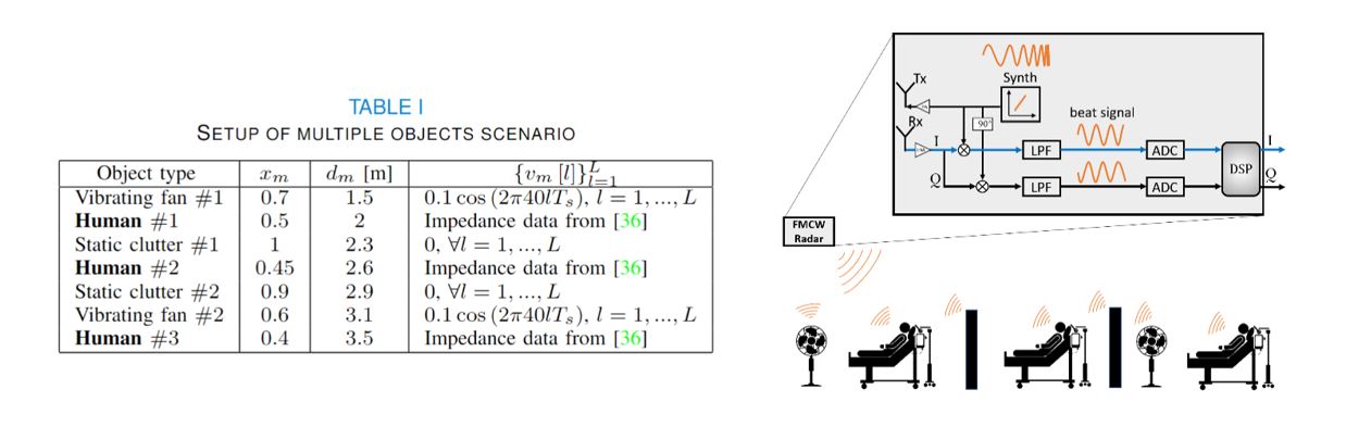
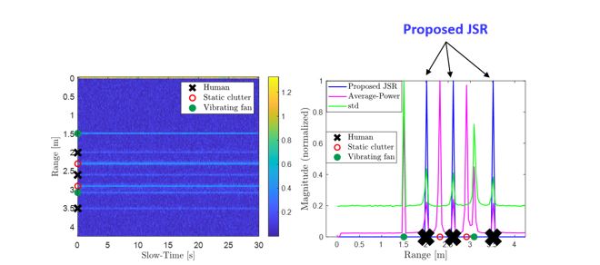
Fig. 2. Multi-person localization by the setup of Table I, for SNR = 0[dB]. (a) The Range vs. Slow-Time map. The row-wise intensities correspond to the DC component and the reflections of Table I 's objects. (b) Several localization techniques based on the average power [4], the std estimate [5] and the proposed JSR [1]. Only JSR localization properly detects the subjects in the given scenario.
Scenario #2:
Multiple objects located at the same radial distance but at different angles – using a SIMO setup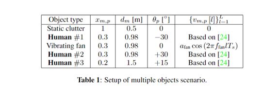
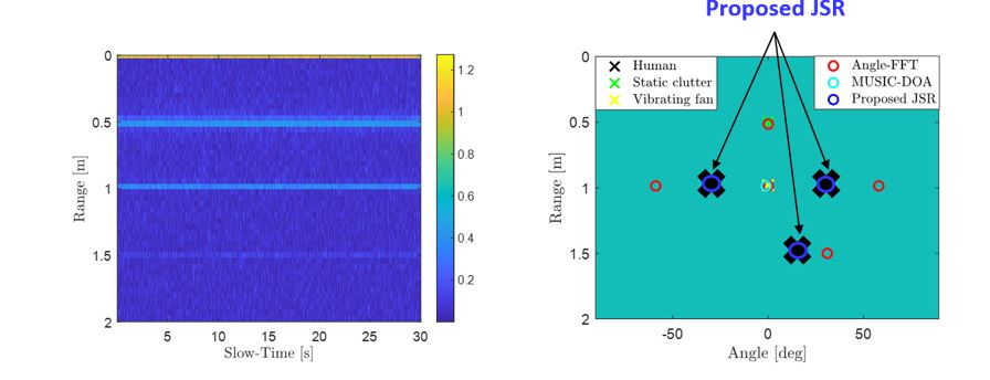
Fig. 3. Multi-person localization by the setup of Table 1 that includes equidistant targets, for SNR=0 [dB]. Only the proposed JSR method properly detects the humans in the given scenario.
B. Multi-Person NCVSM.

Fig. 4. Simultaneous NCVSM of 3 subjects given their thoracic vibrations, w.r.t. known references, SNR=0 [dB].
Rows: 3 different subjects. Columns: Extracted thoracic vibrations for a given time interval, proposed VSDR [1], FFT w/ ZP, FFT w/o ZP and Phase-Reg. The VSDR estimates are the closest to the reference estimates for both RR and HR.
- In terms of performance, the VSDR outperforms the other compared methods in 4 different metrics, for any examined SNR case.
- Specifically, In the more challenging task of HR monitoring, the gap between the VSDR and the other techniques is prominent.
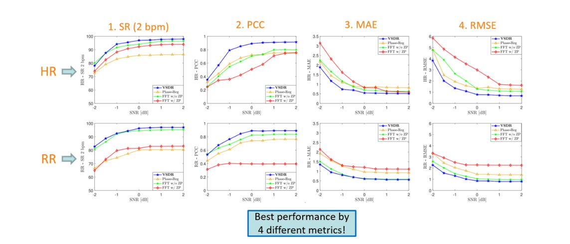
Fig. 5. Median NCVSM performance of the proposed VSDR [1] compared to other techniques vs. SNR. Rows: HR, RR. Columns: SR - 2 bpm, PCC, MAE, RMSE.
Hardware
We developed a dedicated hardware phantom that simulates multi-person thoracic displacements through which realistic experiments for NCVSM using radars can be performed. The phantom reduces the need for human trials and enables proper calibration of the suggested algorithms.
Our demonstration platform consists of (1) a vibration generator for imitating thoracic displacements, (2) flat circular metal plates (24 cm diameter), (3) a radar sensor, located up to 2 meters from the phantom, and (4) a dedicated experimental setup.
Scenario #1:
Hardware phantom for single-person NCVSM via radar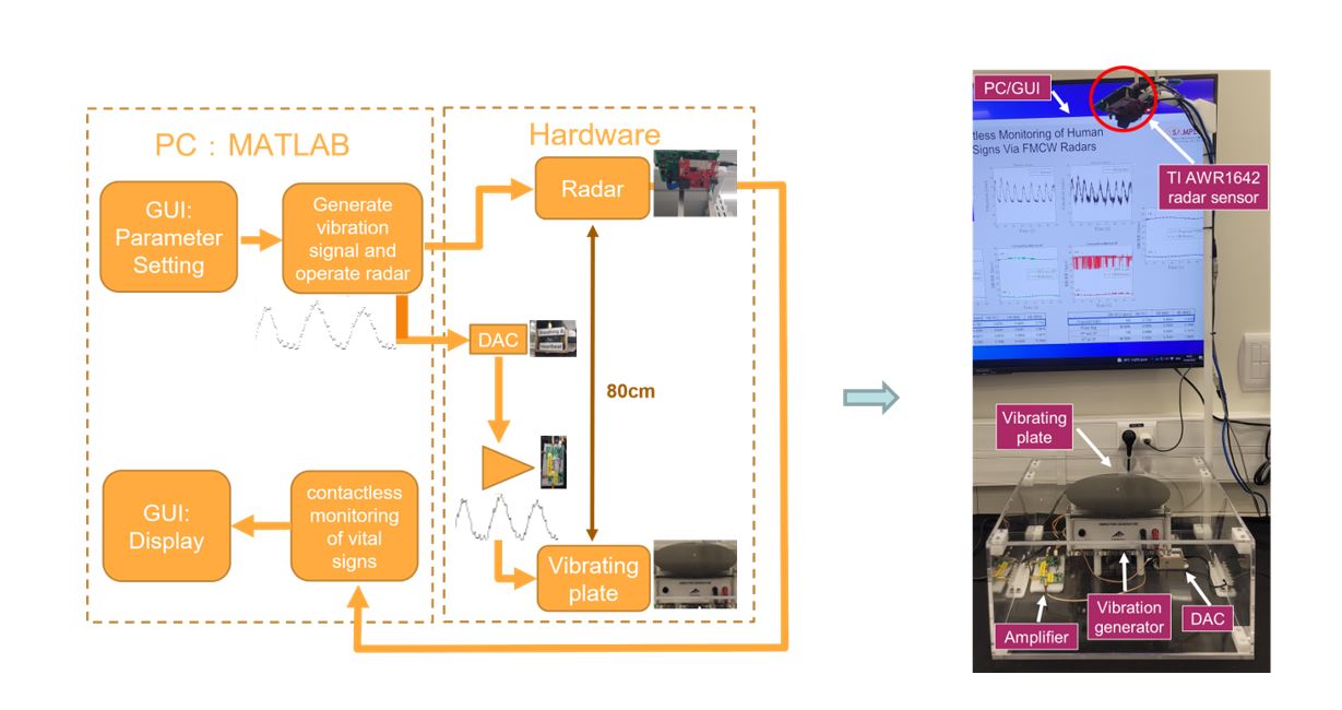
Graphical User Interface:
Scenario #2:
Hardware phantom for multi-person NCVSM via radar.
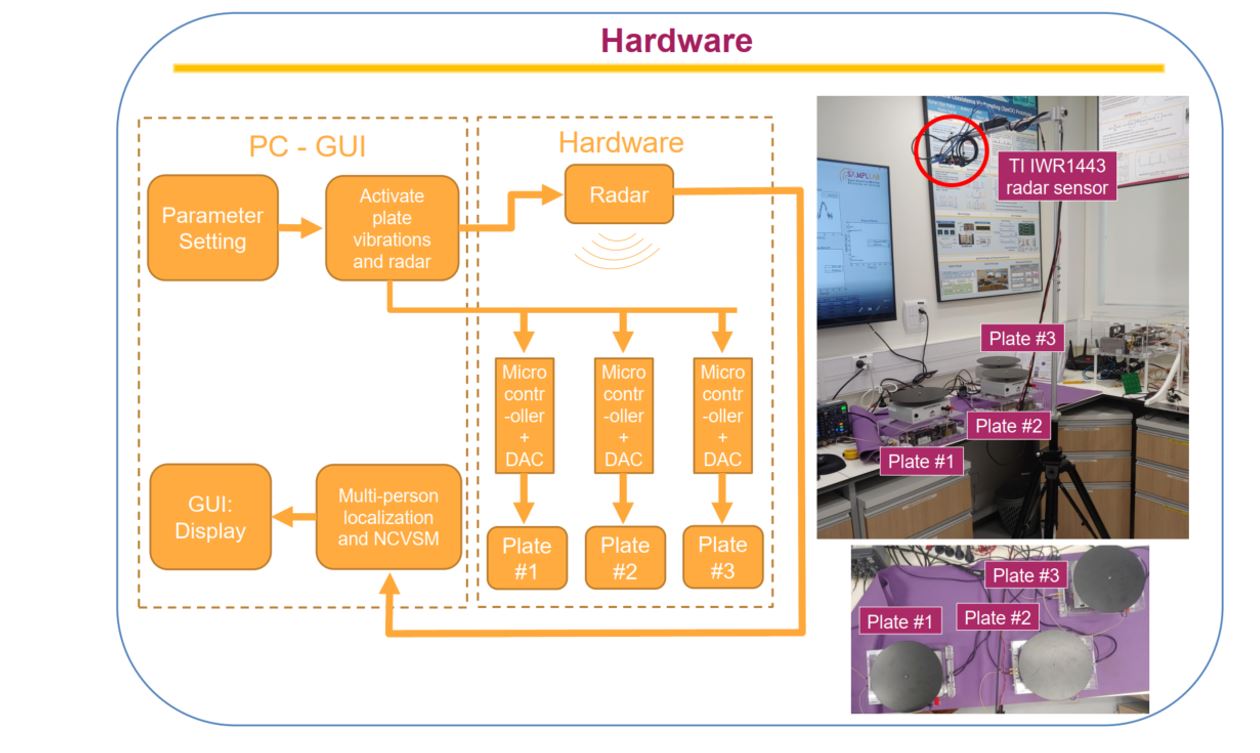
Graphical User Interface
- Example of localization and NCVSM of 3 people simultaneously:
- Two equidistant targets located approximately at 1 [m] from the radar and +-10 [deg]. The third target is located approximately at 1.1 [m] from the radar and +25 [deg] .
- The operator can choose between a trial of sinusoids or human thoracic vibrations as well as determining the frame rate and duration of the trial.
Videos:
ICASSP22 Hardware Demo: Contactless Vital Signs - YouTube.
Sparsity-Based Multi-Person NCVSM Using Radar - YouTube.
ICASSP 2023 IEEE International Conference on Acoustics, Speech and Signal Processing:
Articles from the media
https://www.ynet.co.il/environment-science/article/bytggrhkgg#google_vignette
https://www.israel21c.org/noncontact-radar-unit-monitors-patients-vitals-remotely/
https://www.jpost.com/health-and-wellness/article-853525
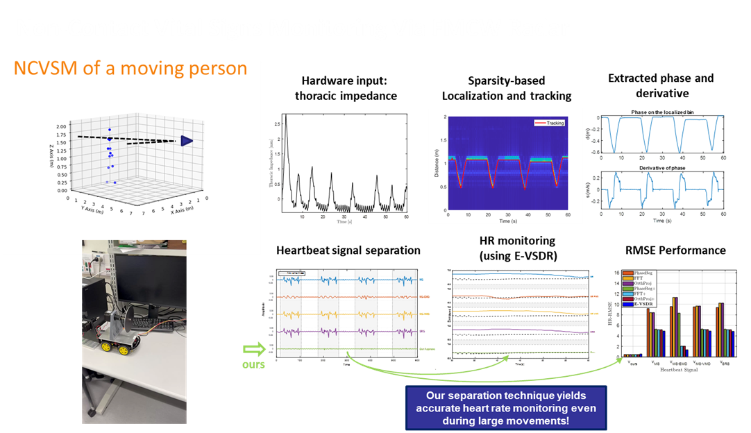
Extension to NCVSM during large movements (such as walking)
- Real-world healthcare scenarios, e.g., patients shifting in bed, or monitoring uncooperative subjects, require robust non-contact vital sign monitoring (NCVSM) during movement
- Proposed solution for moving people using SISO FMCW radar: extends [1] by
- Sparsity-based localization scheme involving Kalman filtering for tracking people during large movements
- Signal separation framework leveraging cardiopulmonary patterns and movement dynamics
- The Extended Vital Signs-based Dictionary Recovery (E-VSDR) [5] method is then used to continuously estimate the heart rate (HR)
- Our signal separation approach yields superior NCVSM results compared to state-of-the-art separation methods.
Video:
RaLU-Net: Deep unfolded radar localization of humans
-
Rising demand for monitoring solutions in crowded environments
-
Detecting and positioning multiple humans with radar is highly affected by resolution limitations
-
This work first provides the iterative localization method: RaLU-JSR
-
exploits joint sparsity and cardiopulmonary properties
-
- Then, it is unfolded into a neural network: RaLU-Net
-
Utilizes the unique structure of the data to enhance accuracy and reduce computational cost
-
-
Achieving high performance with minimal computation in thousands of close-proximity scenarios


Non-contact Spirometry test
- Non-Contact Spirometry Test (NCST) to evaluate pulmonary function
- Eliminates the need for cumbersome equipment
- Facilitates cooperation especially in children
- Interpretable mathematical modelling
- Polynomial relationship and sparse recovery techniques


Non-Invasive Quantitative Monitoring of Nanoparticles In Vivo Using Millimeter Wave Radar
The identification of nanoparticles in specific regions of the body can indicate the presence of pathological conditions such as tumors, metastases, or infections. This occurs either due to the natural tendency of nanoparticles to accumulate in areas with enhanced permeability and retention (EPR effect), or through active targeting mechanisms involving conjugation to specific molecular markers. Additionally, detecting nanoparticle accumulation in targeted organs is particularly relevant given that, on average, only 0.7% of the injected dose reaches solid tumors [Wilhelm et al., 2016]. Therefore, the ability to localize and quantify nanoparticles in vivo—and to monitor changes in their concentration over time—has significant potential as a non-invasive diagnostic and monitoring approach.

References:
- Y. Eder and Y. C. Eldar, "Sparsity-Based Multi-Person Non-Contact Vital Signs Monitoring via FMCW Radar," IEEE Journal of Biomedical and Health Informatics, vol. 27, no. 6, pp. 2806-2817, 2023.
- Y. Eder, Z. Liu, and Y. C. Eldar, "Sparse Non-Contact Multiple People Localization and Vital Signs Monitoring Via FMCW Radar," in Proc. IEEE Int. Conf. Acoust., Speech, Signal Process. (ICASSP), Rhodes Island, Greece, 2023, pp. 1-5.
- Y. Eder, R. Abel, A. Schroeder, and Y. C. Eldar, "Localization and Tracking of Gold Nanoparticles Using mmWave FMCW Radar," in Proc. IEEE Int. Conf. Acoust., Speech, Signal Process. (ICASSP), 2024, pp. 2066-2070.
- Y. Eder, Y. Kvich, R. Guo, and Y. C. Eldar, "RaLU-Net: Deep Unfolded Radar Localization of Humans for Precise Multi-Person Non-Contact Vital Signs Monitoring," in Proc. IEEE Int. Conf. Acoust., Speech, Signal Process. (ICASSP), April 2025, pp. 1-5.
- Y. Eder, E. Zagoury, S. Savariego, M. Namer, O. Cohen, and Y. C. Eldar, "Robust Phantom-Assisted Framework for Multi-Person Localization and Vital Signs Monitoring Using MIMO FMCW Radar," arXiv preprint, arXiv:2501.06755, 2025.