
Atomic force microscopy is best known for its powerful imaging capabilities. Surfaces are imaged with resolution down to the molecular level with capability to access 100 x 100 micron field of view. The instrument can operate in ambient, liquid, and vacuum environments and at over a temperature range from -30 degrees to + 100 degrees C.
Locally-induced crystallization in thermoplastic
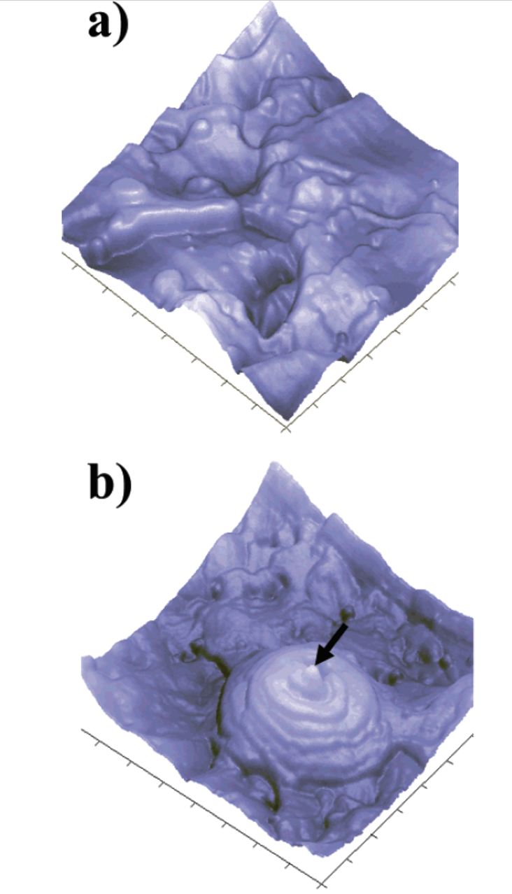
Nanoletters 4, 1439 (2004)
“volcanic pools” Phase image of gold grains

Fibers
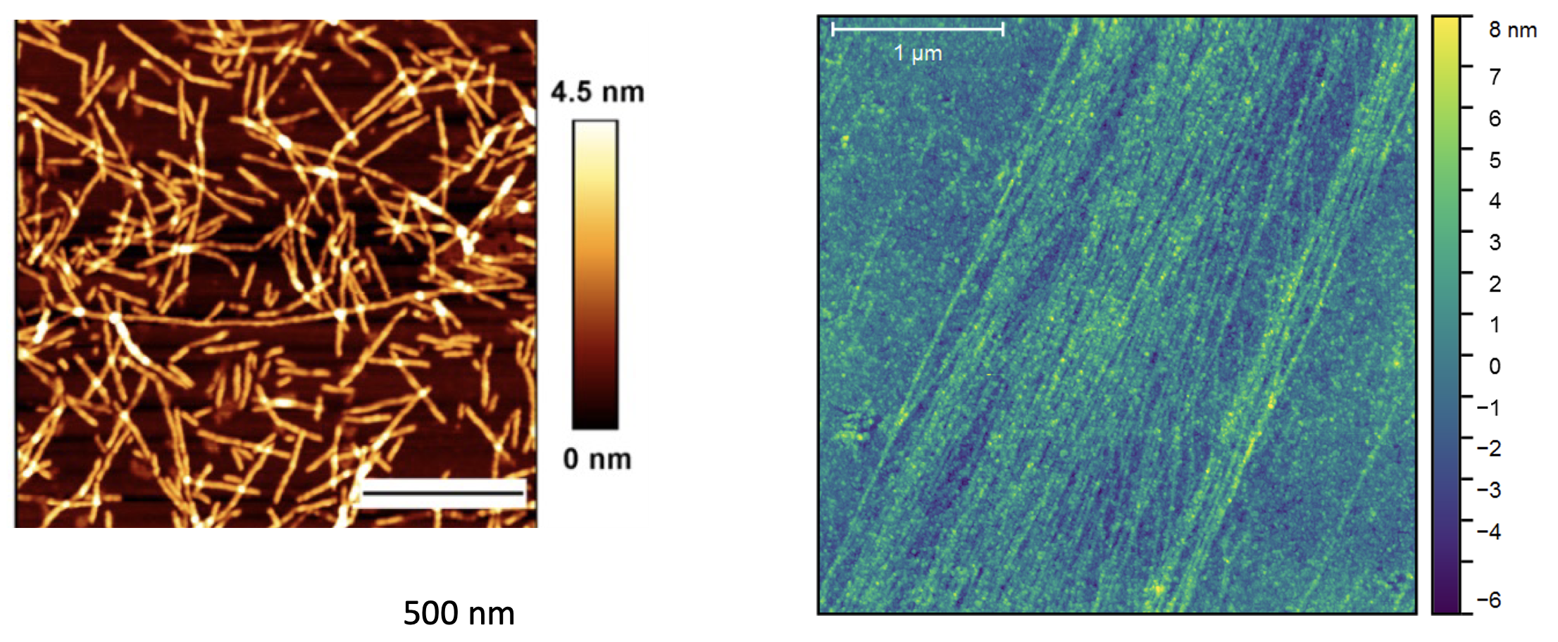
Communications Chemistry 4, 62 (2021)
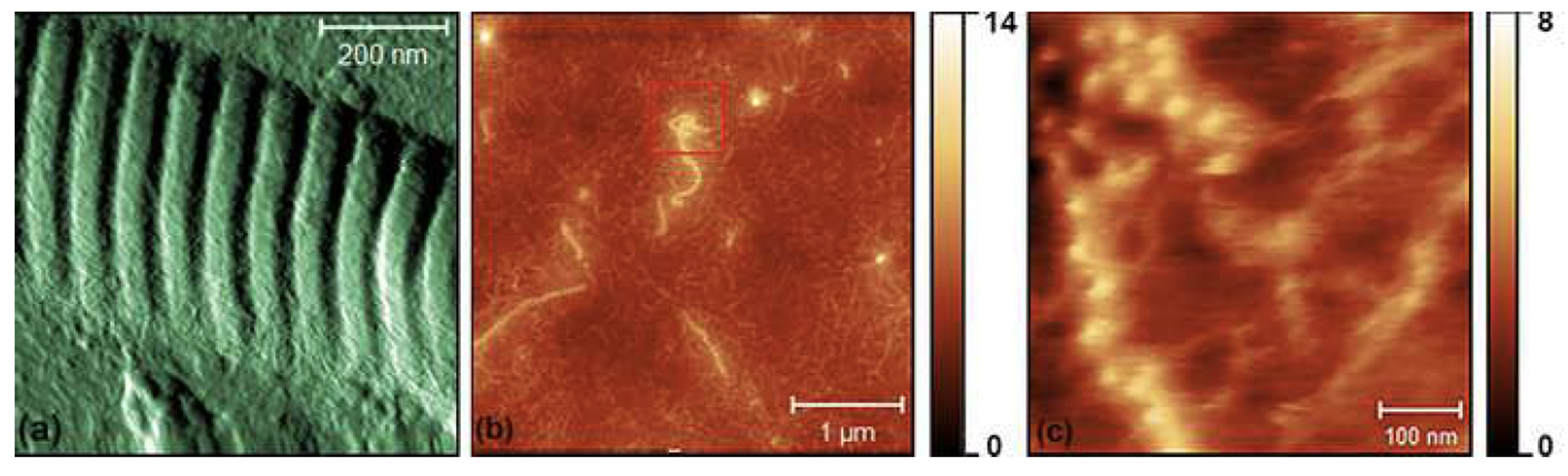
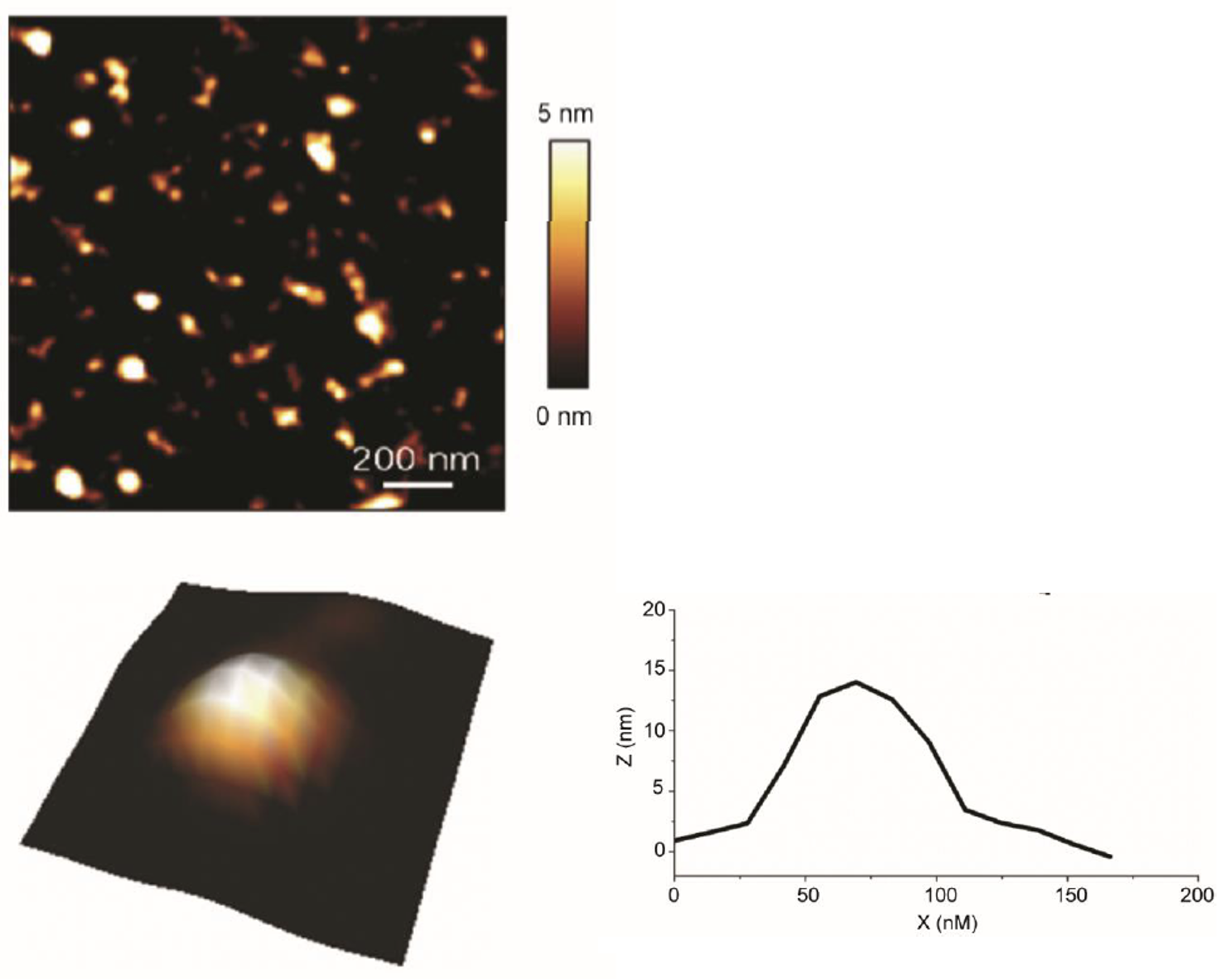
“Brainy” nanoparticles of metal-organic framework

Angew. Chem. Int. Ed. 60, 18256–18264 (2021)
Collagen intact, and enzyme-digested
Isr. J. Chem. 60, 1171 – 1184 (2020)
Imaging Red blood cell cytoskeleton
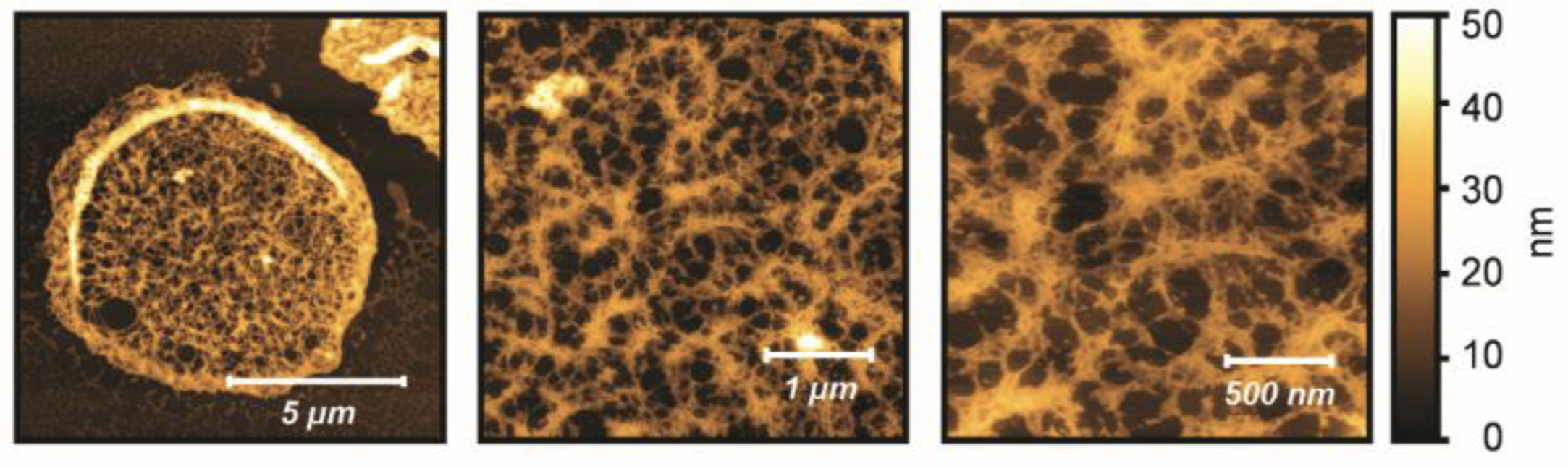
Nature Communications 12:1172 (2021)
Imaging Extracellular vesicles
Nature Communications 12:1172 (2021)
Using supervised machine learning for classification of AFM images
Analysis and classification of RBC cytoskeleton fast-AFM images using supervised machine learning (Convoutional Neural Networks)

Beilstein Journal of Nanotechnology 12, 871-901 (2021)
Measurements of swelling and mechanical properties of carbon micro-inclusions in flint

Chemical Geology 582, 120427 (2021)