
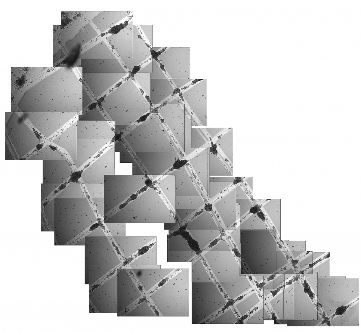
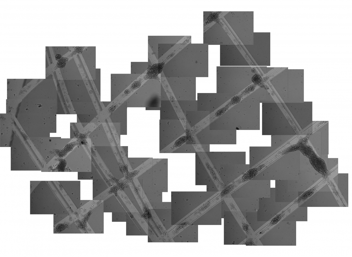
full size full size
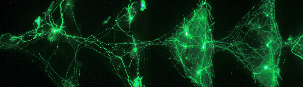
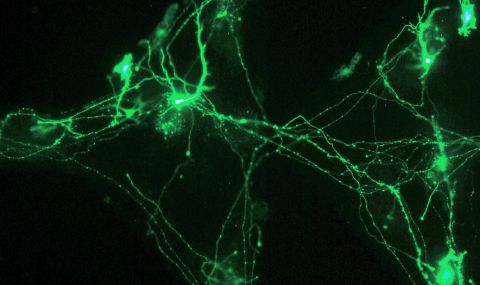
P19 embryonal carcinoma cells differentiated into neuron-like cells, 24 days after plating.
The plated cells stick exclusively on designated lines (seen as the lighter lines in the picture) covered with laminin (sigma L2020).
Within a day after being plated, the cells form neurites along the laminin lines and establish connections. Within the first 3 days neighboring cells will group together to form aggregates, the neurites of cells from the same aggregate stitch together to make thick fascicles which connect neighboring aggregates.
There are in the order of 100 neurons in each of the aggregates seen in these pictures.
Note that some neurites seem to be unrestricted to the line and 'cut corners', this can happen because neurites are often attached to the substrates by their tip only, the sticky tip can follow the corner, while the length of the neurite unattached to the surface can cut the corner.
At the time this picture was taken (24 days after plating), the neurons are electrically active. They exhibit spiking behavior similar to cortical neurons and form functional synapses.
Both pictures are of the same area, Pic. 1 (Left) taken with a 5X lens and Pic. 2 (Right) with a 10X lens. The pictures were taken through a Zeiss Axiovert 135 TV microscope, and COHU CCD. A collage of many frames enables the large field of view shown in the pictures.