Sample preparation
For sample preparation for mass spectrometry imaging the unit offers all necessary equipment:
- cryostat 1950(Leica),
- freeze dryer (Martin Christ),
- vacuum desiccator,
- M3 sprayer (HTXimaging) for matrix application,
- Axio 16 stereoscope (Zeiss)
Furthermore we offer different cryo-sectioning and sample preparation protocols (e.g. cryo-film method, sectioning of embedded tissue, mounting of Tissues on other substrates, etc.) as a service or train our collaborators to prepare the samples in a mass spectrometry compatible way.
One time training of students is mandatory for future acceptance of samples directly for mass spectrometry imaging. 
Figure: Workflow of sample preparation for mass spectrometry imaging, starting from sample collection, sectioning, and desiccation, optional chemical or biochemical modifications, matrix application in case of MALDI imaging and analysis.
Mass spectrometry imaging
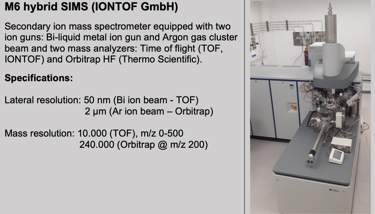
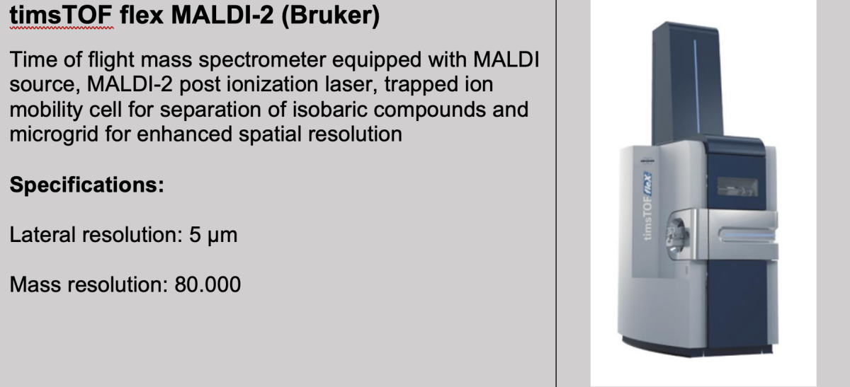
The unit currently operates two high-end imaging mass spectrometers, the M6 hybrid SIMS for secondary ion mass spectrometry imaging and the timsTOF flex MALDI-2 for MALDI imaging.
Data analysis
The unit offers data processing, statistical analysis and visualization with vendors’ software and “home-made” software solutions:
- flexImaging (Bruker, MALDI),
- SCiLSTM lab (Bruker, MALDI),
- SurfaceLabTM (IONTOF, SIMS),
- MALDIquant (R package, MALDI)
- MSiReader (Matlab, MALDI)
- ShinyCardinal (In-house comprehensive analysis software with GUI for MSI analysis)
- Other home-made software (e.g. PICA, Dong et al. 2023)
For users who want to analyze/visualize their data by themselves we provide a workstation in the unit providing full licenses of needed software packages.